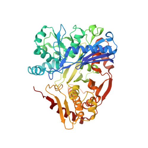
Nucleant-mediated protein crystallization with the application of microporous synthetic zeolites.
Sugahara, M., Asada, Y., Morikawa, Y., Kageyama, Y., Kunishima, N.(2008) Acta Crystallogr D Biol Crystallogr 64: 686-695
- PubMed: 18560157
- DOI: https://doi.org/10.1107/S0907444908009980
- Primary Citation of Related Structures:
1WMM, 2DPN, 2HD9, 2ZBN - PubMed Abstract:
Protein crystallization is still a major bottleneck in structural biology. As the current methodology of protein crystallization is a type of screening, it is usually difficult to crystallize important target proteins. It was thought that hetero-epitaxic growth from the surface of a mineral crystal acting as a nucleant would be an effective enhancer of protein crystallization. However, in spite of almost two decades of effort, a generally applicable hetero-epitaxic nucleant for protein crystallization has yet to be found. Here we introduce the first candidate for a universal hetero-epitaxic nucleant, microporous zeolite: a synthetic aluminosilicate crystalline polymer with regular micropores. It promotes a form-selective crystal nucleation of proteins and acts as a crystallization catalyst. The most successful zeolite nucleant was molecular sieve type 5A with a pore size of 5 A and with bound Ca2+ ions. The zeolite-mediated crystallization improved the crystal quality in five out of six proteins tested. It provided new crystal forms with better resolution in two cases, larger crystals in one case, and zeolite-dependent crystal formations in two cases. The hetero-epitaxic growth of the zeolite-mediated crystals was confirmed by a crystal-packing analysis which revealed a layer-like structure in the crystal lattice.
Organizational Affiliation:
RIKEN SPring-8 Center, Harima Institute, 1-1-1 Kouto, Sayo-cho, Sayo-gun, Hyogo 679-5148, Japan.


