2D05

Chitosanase From Bacillus circulans mutant K218P


Experimental Data Snapshot

  • Method: X-RAY DIFFRACTION
  • Resolution: 2.00 Å
  • R-Value Free: 0.213 
  • R-Value Work: 0.173 

wwPDB Validation   3D Report Full Report


This is version 1.3 of the entry. See complete history


Literature

Bacillus circulans MH-K1 Chitosanase: Amino Acid Residues Responsible for Substrate Binding

Fukamizo, T.Amano, S.Yamaguchi, K.Yoshikawa, T.Katsumi, T.Saito, J.Suzuki, M.Miki, K.Nagata, Y.Ando, A.

(2005) J Biochem 138: 563-569

  • DOI: https://doi.org/10.1093/jb/mvi156
  • Primary Citation of Related Structures:  
    2D05

  • PubMed Abstract: 

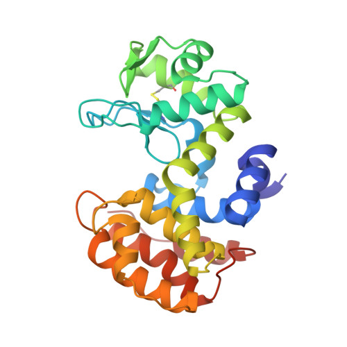
    To identify the amino acids responsible for the substrate binding of chitosanase from Bacillus circulans MH-K1 (MH-K1 chitosanase), Tyr148 and Lys218 of the chitosanase were mutated to serine and proline, respectively, and the mutated chitosanases were characterized. The enzymatic activities of Y148S and K218P were found to be 12.5% and 0.16% of the wild type, respectively. When the (GlcN)3 binding ability to the chitosanase was evaluated by fluorescence spectroscopy and thermal unfolding experiments, the binding abilities of both mutant enzymes were markedly reduced as compared with the wild type enzyme. The affinity of the enzyme for the trisaccharide decreased by 1.0 kcal/mol of binding free energy for Y148S, and 3.7 kcal/mol for K218P. The crystal structure of K218P revealed that Pro218 forms a cis-peptide bond and that the state of the flexible loop containing the 218th residue is considerably affected by the mutation. Thus, we conclude that the flexible loop containing Lys218 plays an important role in substrate binding, and that the role of Tyr148 is less critical, but still important, due to a stacking interaction or hydrogen bond.


  • Organizational Affiliation

    Department of Advanced Bioscience, Kinki University, 3327-204 Nakamachi, Nara 631-8505. fukamizo@nara.kindai.ac.jp


Macromolecules
Find similar proteins by:  (by identity cutoff)  |  3D Structure
Entity ID: 1
MoleculeChains Sequence LengthOrganismDetailsImage
Chitosanase259Niallia circulansMutation(s): 1 
EC: 3.2.1.132
UniProt
Find proteins for P33673 (Niallia circulans)
Explore P33673 
Go to UniProtKB:  P33673
Entity Groups  
Sequence Clusters30% Identity50% Identity70% Identity90% Identity95% Identity100% Identity
UniProt GroupP33673
Sequence Annotations
Expand
  • Reference Sequence
Small Molecules
Ligands 1 Unique
IDChains Name / Formula / InChI Key2D Diagram3D Interactions
SO4
Query on SO4

Download Ideal Coordinates CCD File 
B [auth A]SULFATE ION
O4 S
QAOWNCQODCNURD-UHFFFAOYSA-L
Experimental Data & Validation

Experimental Data

  • Method: X-RAY DIFFRACTION
  • Resolution: 2.00 Å
  • R-Value Free: 0.213 
  • R-Value Work: 0.173 
  • Space Group: P 21 21 2
Unit Cell:
Length ( Å )Angle ( ˚ )
a = 43.3α = 90
b = 128.1β = 90
c = 57.8γ = 90
Software Package:
Software NamePurpose
DENZOdata reduction
SCALEPACKdata scaling
AMoREphasing
X-PLORrefinement

Structure Validation

View Full Validation Report


Entry History 

Deposition Data

Revision History  (Full details and data files)

  • Version 1.0: 2005-12-06
    Type: Initial release
  • Version 1.1: 2008-04-30
    Changes: Version format compliance
  • Version 1.2: 2011-07-13
    Changes: Version format compliance
  • Version 1.3: 2021-11-10
    Changes: Database references, Derived calculations