2CZS

Crystal Structure Analysis of the Diheme c-type Cytochrome DHC2


Experimental Data Snapshot

  • Method: X-RAY DIFFRACTION
  • Resolution: 1.50 Å
  • R-Value Free: 0.272 
  • R-Value Work: 0.221 
  • R-Value Observed: 0.224 

wwPDB Validation   3D Report Full Report


Ligand Structure Quality Assessment 


This is version 1.2 of the entry. See complete history


Literature

Structural and Biochemical Characterization of DHC2, a Novel Diheme Cytochrome c from Geobacter sulfurreducens

Heitmann, D.Einsle, O.

(2005) Biochemistry 44: 12411-12419

  • DOI: https://doi.org/10.1021/bi0509999
  • Primary Citation of Related Structures:  
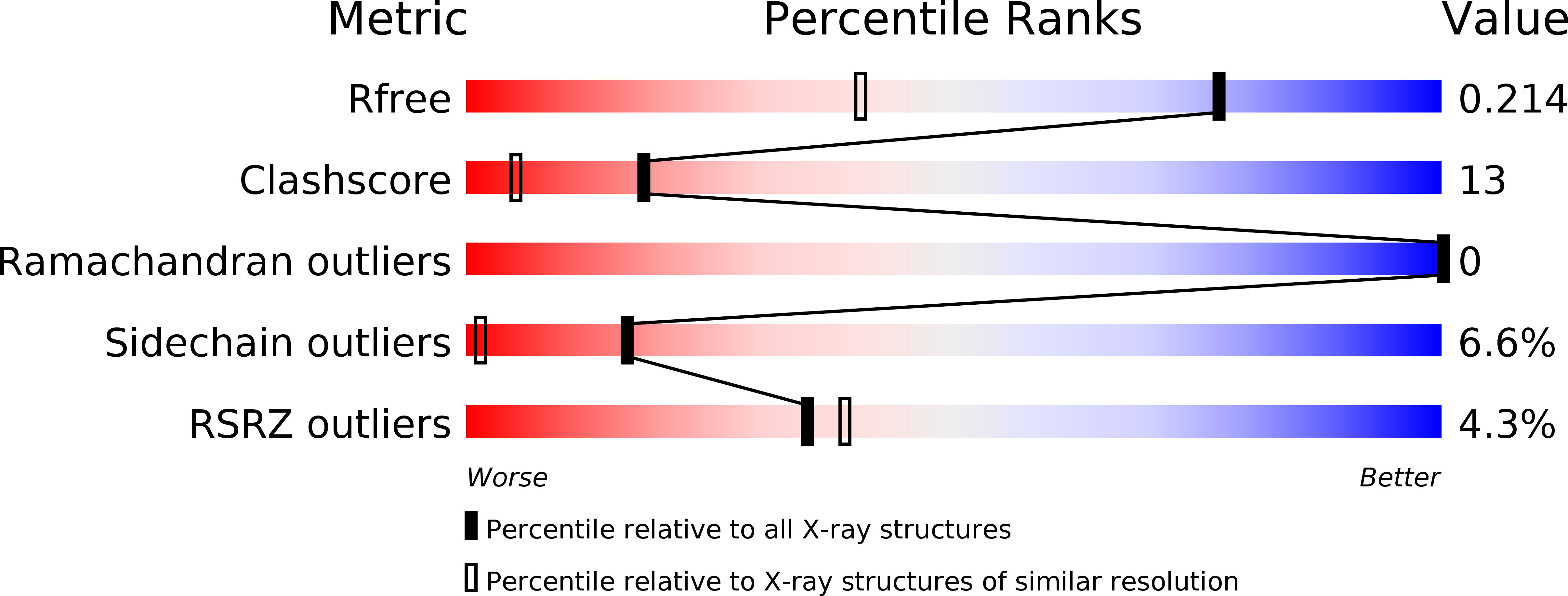
    2CZS

  • PubMed Abstract: 

    Multiheme cytochromes c constitute a widespread class of proteins with essential functions in electron transfer and enzymatic catalysis. Their functional properties are in part determined by the relative arrangement of multiple heme cofactors, which in many cases have been found to pack in conserved interaction motifs. Understanding the significance of these motifs is crucial for the elucidation of the highly optimized properties of multiheme cytochromes c, but their spectroscopic investigation is often hindered by the large number and efficient coupling of the individual centers and the limited availability of recombinant protein material. We have identified a diheme cytochrome c, DHC2, from the metal-reducing soil bacterium Geobacter sulfurreducens and determined its crystal structure by the method of multiple-wavelength anomalous dispersion (MAD). The two heme groups of DHC2 pack into one of the typical heme interaction motifs observed in larger multiheme cytochromes, but because of the absence of further, interfering cofactors, the properties of this heme packing motif can be conveniently studied in detail. Spectroscopic properties (UV-vis and EPR) of the protein are typical for cytochromes containing low-spin Fe(III) centers with bis-histidinyl coordination. Midpoint potentials for the two heme groups have been determined to be -135 and -289 mV by potentiometric redox titrations. DHC2 has been produced by recombinant expression in Escherichia coli using the accessory plasmid pEC86 and is therefore accessible for systematic mutational studies in further investigating the properties of heme packing interactions in cytochromes c.


  • Organizational Affiliation

    Institut für Mikrobiologie und Genetik, Abteilung Molekulare Strukturbiologie, Georg-August-Universität Göttingen, Justus-von-Liebig-Weg 11, 37077 Göttingen, Germany.


Macromolecules
Find similar proteins by:  (by identity cutoff)  |  3D Structure
Entity ID: 1
MoleculeChains Sequence LengthOrganismDetailsImage
cytochrome c, putative
A, B
80Geobacter sulfurreducens PCAMutation(s): 0 
UniProt
Find proteins for Q748S4 (Geobacter sulfurreducens (strain ATCC 51573 / DSM 12127 / PCA))
Explore Q748S4 
Go to UniProtKB:  Q748S4
Entity Groups  
Sequence Clusters30% Identity50% Identity70% Identity90% Identity95% Identity100% Identity
UniProt GroupQ748S4
Sequence Annotations
Expand
  • Reference Sequence
Small Molecules
Ligands 3 Unique
IDChains Name / Formula / InChI Key2D Diagram3D Interactions
HEM
Query on HEM

Download Ideal Coordinates CCD File 
F [auth A],
G [auth A],
K [auth B],
L [auth B]
PROTOPORPHYRIN IX CONTAINING FE
C34 H32 Fe N4 O4
KABFMIBPWCXCRK-RGGAHWMASA-L
SO4
Query on SO4

Download Ideal Coordinates CCD File 
C [auth A],
D [auth A],
H [auth B],
I [auth B]
SULFATE ION
O4 S
QAOWNCQODCNURD-UHFFFAOYSA-L
NA
Query on NA

Download Ideal Coordinates CCD File 
E [auth A],
J [auth B]
SODIUM ION
Na
FKNQFGJONOIPTF-UHFFFAOYSA-N
Experimental Data & Validation

Experimental Data

  • Method: X-RAY DIFFRACTION
  • Resolution: 1.50 Å
  • R-Value Free: 0.272 
  • R-Value Work: 0.221 
  • R-Value Observed: 0.224 
  • Space Group: P 1 21 1
Unit Cell:
Length ( Å )Angle ( ˚ )
a = 39.645α = 90
b = 55.679β = 105.91
c = 39.637γ = 90
Software Package:
Software NamePurpose
REFMACrefinement
DENZOdata reduction
SCALEPACKdata scaling

Structure Validation

View Full Validation Report


Ligand Structure Quality Assessment 


Entry History 

Deposition Data

Revision History  (Full details and data files)

  • Version 1.0: 2005-10-18
    Type: Initial release
  • Version 1.1: 2008-04-30
    Changes: Version format compliance
  • Version 1.2: 2011-07-13
    Changes: Derived calculations, Source and taxonomy, Version format compliance