2CW9

Crystal structure of human Tim44 C-terminal domain


Experimental Data Snapshot

  • Method: X-RAY DIFFRACTION
  • Resolution: 1.90 Å
  • R-Value Free: 0.221 
  • R-Value Work: 0.218 
  • R-Value Observed: 0.218 

wwPDB Validation   3D Report Full Report


This is version 1.2 of the entry. See complete history


Literature

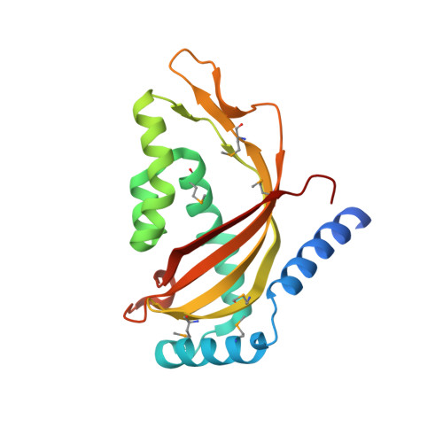
Structure of the human Tim44 C-terminal domain in complex with pentaethylene glycol: ligand-bound form.

Handa, N.Kishishita, S.Morita, S.Akasaka, R.Jin, Z.Chrzas, J.Chen, L.Liu, Z.J.Wang, B.C.Sugano, S.Tanaka, A.Terada, T.Shirouzu, M.Yokoyama, S.

(2007) Acta Crystallogr D Biol Crystallogr 63: 1225-1234

  • DOI: https://doi.org/10.1107/S0907444907051463
  • Primary Citation of Related Structures:  
    2CW9

  • PubMed Abstract: 

    Familial oncocytic thyroid carcinoma is associated with a missense mutation, P308Q, in the C-terminal domain of Tim44. Tim44 is the mitochondrial inner-membrane translocase subunit and it functions as a membrane anchor for the mitochondrial heat-shock protein 70 (mtHsp70). Here, the crystal structure of the human Tim44 C-terminal domain complexed with pentaethylene glycol has been determined at 1.9 A resolution. The overall structure resembles that of the nuclear transport factor 2-like domain. In the crystal structure, pentaethylene glycol molecules are associated at two potential membrane-binding sites: the large hydrophobic cavity and the highly conserved loop between the alpha1 and alpha2 helices near Pro308. A comparison with the yeast homolog revealed that lipid binding induces conformational changes around the alpha1-alpha2 loop, leading to slippage of the alpha1 helix along the large beta-sheet. These changes may play important roles in the translocation of polypeptides across the mitochondrial inner membrane.


  • Organizational Affiliation

    Protein Research Group, Genomic Sciences Center, Yokohama Institute, RIKEN, 1-7-22 Suehiro, Tsurumi, Yokohama 230-0045, Japan.


Macromolecules
Find similar proteins by:  (by identity cutoff)  |  3D Structure
Entity ID: 1
MoleculeChains Sequence LengthOrganismDetailsImage
translocase of inner mitochondrial membrane194Homo sapiensMutation(s): 0 
UniProt & NIH Common Fund Data Resources
Find proteins for O43615 (Homo sapiens)
Explore O43615 
Go to UniProtKB:  O43615
PHAROS:  O43615
GTEx:  ENSG00000104980 
Entity Groups  
Sequence Clusters30% Identity50% Identity70% Identity90% Identity95% Identity100% Identity
UniProt GroupO43615
Sequence Annotations
Expand
  • Reference Sequence
Small Molecules
Modified Residues  1 Unique
IDChains TypeFormula2D DiagramParent
MSE
Query on MSE
A
L-PEPTIDE LINKINGC5 H11 N O2 SeMET
Experimental Data & Validation

Experimental Data

  • Method: X-RAY DIFFRACTION
  • Resolution: 1.90 Å
  • R-Value Free: 0.221 
  • R-Value Work: 0.218 
  • R-Value Observed: 0.218 
  • Space Group: P 65 2 2
Unit Cell:
Length ( Å )Angle ( ˚ )
a = 108.119α = 90
b = 108.119β = 90
c = 114.056γ = 120
Software Package:
Software NamePurpose
CNSrefinement
HKL-2000data reduction
SCALEPACKdata scaling
SOLVEphasing

Structure Validation

View Full Validation Report


Entry History 

Revision History  (Full details and data files)

  • Version 1.0: 2005-12-17
    Type: Initial release
  • Version 1.1: 2007-10-30
    Changes: Version format compliance
  • Version 1.2: 2011-07-13
    Changes: Version format compliance