2CJM

Mechanism of CDK inhibition by active site phosphorylation: CDK2 Y15p T160p in complex with cyclin A structure


Experimental Data Snapshot

  • Method: X-RAY DIFFRACTION
  • Resolution: 2.30 Å
  • R-Value Free: 0.265 
  • R-Value Work: 0.201 
  • R-Value Observed: 0.204 

wwPDB Validation   3D Report Full Report


Ligand Structure Quality Assessment 


This is version 1.2 of the entry. See complete history


Literature

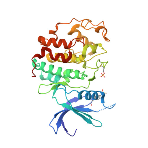
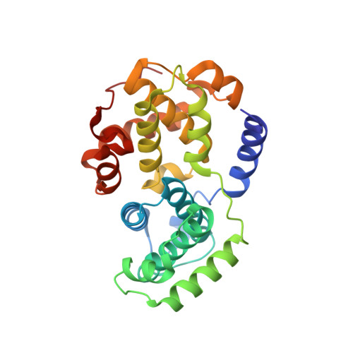
How Tyrosine 15 Phosphorylation Inhibits the Activity of Cyclin-Dependent Kinase 2-Cyclin A.

Welburn, J.P.I.Tucker, J.Johnson, T.Lindert, L.Morgan, M.Willis, A.Noble, M.E.M.Endicott, J.A.

(2007) J Biol Chem 282: 3173

  • DOI: https://doi.org/10.1074/jbc.M609151200
  • Primary Citation of Related Structures:  
    2CJM

  • PubMed Abstract: 

    Inhibition of cyclin-dependent kinase 1 (CDK1) activity by Tyr-15 phosphorylation directly regulates entry into mitosis and is an important element in the control of the unperturbed cell cycle. Active site phosphorylation of other members of the CDK family that regulate cell cycle progression instates checkpoints that are fundamental to eukaryotic cell cycle regulation. Kinetic and crystallographic analyses of CDK2-cyclin A complexes reveal that this inhibitory mechanism operates through steric blockade of peptide substrate binding and through the creation of an environment that favors a non-productive conformation of the terminal group of ATP. By contrast, tyrosine phosphorylation of CDK2 alters neither its Km for ATP nor its significant intrinsic ATPase activity. Tyr-15-phosphorylated CDK2 retains trace protein phosphorylation activity that should be considered in quantitative and qualitative cell cycle models.


  • Organizational Affiliation

    AstraZeneca Pharmaceuticals, Alderley Park, Macclesfield, Cheshire SK10 4TF, United Kingdom.


Macromolecules
Find similar proteins by:  (by identity cutoff)  |  3D Structure
Entity ID: 1
MoleculeChains Sequence LengthOrganismDetailsImage
CELL DIVISION PROTEIN KINASE 2
A, C
298Homo sapiensMutation(s): 0 
EC: 2.7.1.37
UniProt & NIH Common Fund Data Resources
Find proteins for P24941 (Homo sapiens)
Explore P24941 
Go to UniProtKB:  P24941
PHAROS:  P24941
GTEx:  ENSG00000123374 
Entity Groups  
Sequence Clusters30% Identity50% Identity70% Identity90% Identity95% Identity100% Identity
UniProt GroupP24941
Sequence Annotations
Expand
  • Reference Sequence
Find similar proteins by:  (by identity cutoff)  |  3D Structure
Entity ID: 2
MoleculeChains Sequence LengthOrganismDetailsImage
CYCLIN A2
B, D
258Homo sapiensMutation(s): 0 
UniProt & NIH Common Fund Data Resources
Find proteins for P20248 (Homo sapiens)
Explore P20248 
Go to UniProtKB:  P20248
PHAROS:  P20248
GTEx:  ENSG00000145386 
Entity Groups  
Sequence Clusters30% Identity50% Identity70% Identity90% Identity95% Identity100% Identity
UniProt GroupP20248
Sequence Annotations
Expand
  • Reference Sequence
Small Molecules
Modified Residues  2 Unique
IDChains TypeFormula2D DiagramParent
PTR
Query on PTR
A, C
L-PEPTIDE LINKINGC9 H12 N O6 PTYR
TPO
Query on TPO
A, C
L-PEPTIDE LINKINGC4 H10 N O6 PTHR
Experimental Data & Validation

Experimental Data

  • Method: X-RAY DIFFRACTION
  • Resolution: 2.30 Å
  • R-Value Free: 0.265 
  • R-Value Work: 0.201 
  • R-Value Observed: 0.204 
  • Space Group: P 21 21 21
Unit Cell:
Length ( Å )Angle ( ˚ )
a = 73.801α = 90
b = 133.202β = 90
c = 147.585γ = 90
Software Package:
Software NamePurpose
REFMACrefinement
MOSFLMdata reduction
SCALAdata scaling
MOLREPphasing

Structure Validation

View Full Validation Report


Ligand Structure Quality Assessment 


Entry History 

Deposition Data

Revision History  (Full details and data files)

  • Version 1.0: 2006-04-24
    Type: Initial release
  • Version 1.1: 2013-07-10
    Changes: Atomic model, Database references, Derived calculations, Non-polymer description, Other, Source and taxonomy, Structure summary, Version format compliance
  • Version 1.2: 2023-12-13
    Changes: Data collection, Database references, Derived calculations, Other, Refinement description