2BW3

Three-dimensional structure of the Hermes DNA transposase


Experimental Data Snapshot

  • Method: X-RAY DIFFRACTION
  • Resolution: 2.00 Å
  • R-Value Free: 0.211 
  • R-Value Work: 0.185 

wwPDB Validation   3D Report Full Report


This is version 1.2 of the entry. See complete history


Literature

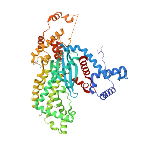
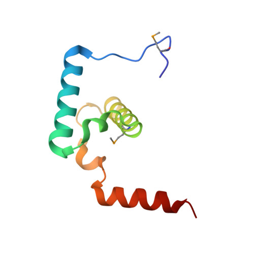
Molecular Architecture of a Eukaryotic DNA Transposase

Hickman, A.B.Perez, Z.N.Zhou, L.Musingarimi, P.Ghirlando, R.Hinshaw, J.E.Craig, N.L.Dyda, F.

(2005) Nat Struct Mol Biol 12: 715

  • DOI: https://doi.org/10.1038/nsmb970
  • Primary Citation of Related Structures:  
    2BW3

  • PubMed Abstract: 

    Mobile elements and their inactive remnants account for large proportions of most eukaryotic genomes, where they have had central roles in genome evolution. Over 50 years ago, McClintock reported a form of stress-induced genome instability in maize in which discrete DNA segments move between chromosomal locations. Our current mechanistic understanding of enzymes catalyzing transposition is largely limited to prokaryotic transposases. The Hermes transposon from the housefly is part of the eukaryotic hAT superfamily that includes hobo from Drosophila, McClintock's maize Activator and Tam3 from snapdragon. We report here the three-dimensional structure of a functionally active form of the transposase from Hermes at 2.1-A resolution. The Hermes protein has some structural features of prokaryotic transposases, including a domain with a retroviral integrase fold. However, this domain is disrupted by the insertion of an additional domain. Finally, transposition is observed only when Hermes assembles into a hexamer.


  • Organizational Affiliation

    Laboratory of Molecular Biology, National Institute of Diabetes and Digestive and Kidney Diseases, National Institutes of Health, Bethesda, Maryland 20892, USA.


Macromolecules
Find similar proteins by:  (by identity cutoff)  |  3D Structure
Entity ID: 1
MoleculeChains Sequence LengthOrganismDetailsImage
TRANSPOSASE534Musca domesticaMutation(s): 3 
UniProt
Find proteins for Q25438 (Musca domestica)
Explore Q25438 
Go to UniProtKB:  Q25438
Entity Groups  
Sequence Clusters30% Identity50% Identity70% Identity90% Identity95% Identity100% Identity
UniProt GroupQ25438
Sequence Annotations
Expand
  • Reference Sequence
Find similar proteins by:  (by identity cutoff)  |  3D Structure
Entity ID: 2
MoleculeChains Sequence LengthOrganismDetailsImage
TRANSPOSASE84Musca domesticaMutation(s): 0 
UniProt
Find proteins for Q25442 (Musca domestica)
Explore Q25442 
Go to UniProtKB:  Q25442
Entity Groups  
Sequence Clusters30% Identity50% Identity70% Identity90% Identity95% Identity100% Identity
UniProt GroupQ25442
Sequence Annotations
Expand
  • Reference Sequence
Small Molecules
Modified Residues  1 Unique
IDChains TypeFormula2D DiagramParent
MSE
Query on MSE
A
L-PEPTIDE LINKINGC5 H11 N O2 SeMET
Experimental Data & Validation

Experimental Data

  • Method: X-RAY DIFFRACTION
  • Resolution: 2.00 Å
  • R-Value Free: 0.211 
  • R-Value Work: 0.185 
  • Space Group: C 1 2 1
Unit Cell:
Length ( Å )Angle ( ˚ )
a = 116.353α = 90
b = 84.974β = 93.71
c = 73.856γ = 90
Software Package:
Software NamePurpose
CNXrefinement
HKL-2000data reduction
SCALEPACKdata scaling
PHASESphasing

Structure Validation

View Full Validation Report


Entry History 

Deposition Data

Revision History  (Full details and data files)

  • Version 1.0: 2005-07-28
    Type: Initial release
  • Version 1.1: 2011-05-08
    Changes: Version format compliance
  • Version 1.2: 2011-07-13
    Changes: Version format compliance