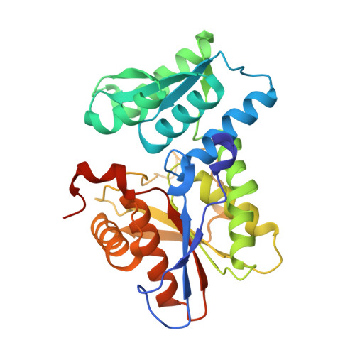
2BHS

Crystal Structure of Cysteine Synthase B


Experimental Data Snapshot

  • Method: X-RAY DIFFRACTION
  • Resolution: 2.67 Å
  • R-Value Free: 0.253 
  • R-Value Work: 0.222 
  • R-Value Observed: 0.223 

wwPDB Validation   3D Report Full Report


Ligand Structure Quality Assessment 


This is version 1.2 of the entry. See complete history


Literature

Structure of the O-Acetylserine Sulfhydrylase Isoenzyme Cysm from Escherichia Coli

Claus, M.T.Zocher, G.E.Maier, T.H.P.Schulz, G.E.

(2005) Biochemistry 44: 8620

  • DOI: https://doi.org/10.1021/bi050485+
  • Primary Citation of Related Structures:  
    2BHS, 2BHT

  • PubMed Abstract: 

    The enzyme O-acetylserine sulfhydrylase participates in the biosynthesis of l-cysteine in bacteria and plants. The structure of isoenzyme B (CysM) from Escherichia coli was established in a hexagonal crystal form at 2.7 A resolution (wild-type) and in a merohedrally twinned tetragonal crystal form at 2.1 A resolution (surface mutant). Structural superpositions revealed the variations with respect to isoenzyme A (CysK) and explained the different substrate specificities. A geometric model of the reaction catalyzed by CysM is proposed. Both isoenzymes are used for the production of l-amino acid derivatives as building blocks for the synthesis of peptides and peptidomimetic drugs. Since the structure of CysM revealed a remarkable main chain variation at the active center, it constitutes a further starting point for engineering mutants with novel substrate specificities.


  • Organizational Affiliation

    Institut für Organische Chemie und Biochemie, Albert-Ludwigs-Universität, Albertstrasse 21, D-79104 Freiburg im Breisgau, Germany.


Macromolecules
Find similar proteins by:  (by identity cutoff)  |  3D Structure
Entity ID: 1
MoleculeChains Sequence LengthOrganismDetailsImage
CYSTEINE SYNTHASE B
A, B, C, D
303Escherichia coliMutation(s): 0 
EC: 2.5.1.47
UniProt
Find proteins for P16703 (Escherichia coli (strain K12))
Explore P16703 
Go to UniProtKB:  P16703
Entity Groups  
Sequence Clusters30% Identity50% Identity70% Identity90% Identity95% Identity100% Identity
UniProt GroupP16703
Sequence Annotations
Expand
  • Reference Sequence
Experimental Data & Validation

Experimental Data

  • Method: X-RAY DIFFRACTION
  • Resolution: 2.67 Å
  • R-Value Free: 0.253 
  • R-Value Work: 0.222 
  • R-Value Observed: 0.223 
  • Space Group: P 65 2 2
Unit Cell:
Length ( Å )Angle ( ˚ )
a = 195.584α = 90
b = 195.584β = 90
c = 235.752γ = 120
Software Package:
Software NamePurpose
REFMACrefinement
XDSdata reduction
XSCALEdata scaling

Structure Validation

View Full Validation Report


Ligand Structure Quality Assessment 


Entry History 

Deposition Data

Revision History  (Full details and data files)

  • Version 1.0: 2005-06-22
    Type: Initial release
  • Version 1.1: 2011-05-08
    Changes: Version format compliance
  • Version 1.2: 2011-07-13
    Changes: Version format compliance