High-resolution structure of calcium-loaded calbindin D9k.
Kordel, J., Skelton, N.J., Akke, M., Chazin, W.J.(1993) J Mol Biol 231: 711-734
- PubMed: 8515447
- DOI: https://doi.org/10.1006/jmbi.1993.1322
- Primary Citation of Related Structures:
2BCA, 2BCB - PubMed Abstract:
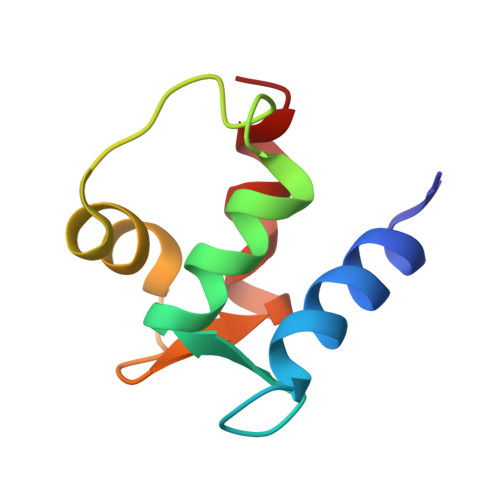
The three-dimensional solution structure of calcium-loaded calbindin D9k has been determined using experimental constraints obtained from nuclear magnetic resonance spectroscopy. A total of 1176 constraints (16 per residue overall, 32 per residue for the core residues) was used for the final refinement, including 1002 distance and 174 dihedral angle constraints. In addition, 23 hydrogen bond constraints were used for the generation of initial structures. Stereospecific assignments were made for 37 of 61 (61%) prochiral methylene protons and the methyl groups of all three valine residues and five out of 12 leucine residues. These constraints were used as input for a series of calculations of three-dimensional structures using a combination of distance geometry and restrained molecular dynamics. The 33 best structures selected for further analysis have no distance constraint violations greater than 0.3 A and good local geometries as reflected by low total energies (< or = -1014 kcal/mol in the AMBER 4.0 force field). The core of the protein consists of four well-defined helices with root-mean-square deviations from the average of 0.45 A for the N, C alpha and C' backbone atoms. These helices are packed in an antiparallel fashion to form two helix-loop-helix calcium-binding motifs, termed EF-hands. The two EF-hands are joined at one end by a ten-residue linker segment, and at the other by a short beta-type interaction between the two calcium-binding loops. Overall, the average solution structure of calbindin D9k is very similar to the crystal structure, with a pairwise root-mean-square deviation of 0.85 A for the N, C alpha and C' backbone atoms of the four helices. The differences that are observed between the solution and the crystal structures are attributed to specific crystal contacts, increased side-chain flexibility in solution, or artifacts arising from molecular dynamics refinement of the solution structures in vacuo.
Organizational Affiliation:
Department of Molecular Biology, Scripps Research Institute La Jolla, CA 92037.