2B18

N-terminal GAF domain of transcriptional pleiotropic repressor CodY.


Experimental Data Snapshot

  • Method: X-RAY DIFFRACTION
  • Resolution: 1.80 Å
  • R-Value Free: 0.212 
  • R-Value Work: 0.154 
  • R-Value Observed: 0.156 

wwPDB Validation   3D Report Full Report


This is version 1.4 of the entry. See complete history


Literature

The Structure of CodY, a GTP- and Isoleucine-responsive Regulator of Stationary Phase and Virulence in Gram-positive Bacteria.

Levdikov, V.M.Blagova, E.Joseph, P.Sonenshein, A.L.Wilkinson, A.J.

(2006) J Biol Chem 281: 11366-11373

  • DOI: https://doi.org/10.1074/jbc.M513015200
  • Primary Citation of Related Structures:  
    2B0L, 2B18, 2GX5, 2HGV

  • PubMed Abstract: 

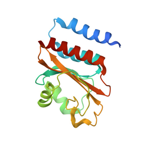
    CodY is a global regulator of transcription in gram-positive bacteria. It represses during growth genes required for adaptation to nutrient limitation, including virulence genes in some human pathogens. CodY activity is regulated by GTP and branched chain amino acids, metabolites whose intracellular concentrations drop as cells enter stationary phase. Although CodY has a highly conserved sequence, it has no significant similarity to proteins of known structure. Here we report crystal structures of two fragments of CodY from Bacillus subtilis that clearly constitute its cofactor and DNA binding domains and reveal that CodY is a chimera of previously observed folding units. The N-terminal cofactor-binding fragment adopts a fold reminiscent of the GAF domains found in cyclic nucleotide phosphodiesterases and adenylate cyclases. It is a dimer stabilized by an intermolecular six alpha-helical bundle that buries an extensive apolar surface rich in residues invariant in CodY orthologues. The branched chain amino acid ligands reside in hydrophobic pockets of each monomer distal to the dimer-forming surface. The structure of the C-terminal DNA binding domain belongs to the winged helix-turn-helix family. The implications of the structure for DNA binding by CodY and its control by cofactor binding are discussed.


  • Organizational Affiliation

    Structural Biology Laboratory, Department of Chemistry, University of York, York YO10 5YW, United Kingdom.


Macromolecules
Find similar proteins by:  (by identity cutoff)  |  3D Structure
Entity ID: 1
MoleculeChains Sequence LengthOrganismDetailsImage
GTP-sensing transcriptional pleiotropic repressor codY164Bacillus subtilisMutation(s): 0 
Gene Names: codY
UniProt
Find proteins for P39779 (Bacillus subtilis (strain 168))
Explore P39779 
Go to UniProtKB:  P39779
Entity Groups  
Sequence Clusters30% Identity50% Identity70% Identity90% Identity95% Identity100% Identity
UniProt GroupP39779
Sequence Annotations
Expand
  • Reference Sequence
Small Molecules
Ligands 1 Unique
IDChains Name / Formula / InChI Key2D Diagram3D Interactions
ILE
Query on ILE

Download Ideal Coordinates CCD File 
B [auth A]ISOLEUCINE
C6 H13 N O2
AGPKZVBTJJNPAG-WHFBIAKZSA-N
Experimental Data & Validation

Experimental Data

  • Method: X-RAY DIFFRACTION
  • Resolution: 1.80 Å
  • R-Value Free: 0.212 
  • R-Value Work: 0.154 
  • R-Value Observed: 0.156 
  • Space Group: P 21 21 2
Unit Cell:
Length ( Å )Angle ( ˚ )
a = 34.696α = 90
b = 86.081β = 90
c = 54.819γ = 90
Software Package:
Software NamePurpose
REFMACrefinement
HKL-2000data reduction
SCALEPACKdata scaling
HKL2Mapphasing

Structure Validation

View Full Validation Report


Entry History 

Deposition Data

Revision History  (Full details and data files)

  • Version 1.0: 2006-02-21
    Type: Initial release
  • Version 1.1: 2008-05-01
    Changes: Version format compliance
  • Version 1.2: 2011-07-13
    Changes: Version format compliance
  • Version 1.3: 2017-10-11
    Changes: Refinement description
  • Version 1.4: 2024-02-14
    Changes: Data collection, Database references, Derived calculations