NMR structure and molecular dynamics of the in-plane membrane anchor of nonstructural protein 5A from bovine viral diarrhea virus.
Sapay, N., Montserret, R., Chipot, C., Brass, V., Moradpour, D., Deleage, G., Penin, F.(2006) Biochemistry 45: 2221-2233
- PubMed: 16475810
- DOI: https://doi.org/10.1021/bi0517685
- Primary Citation of Related Structures:
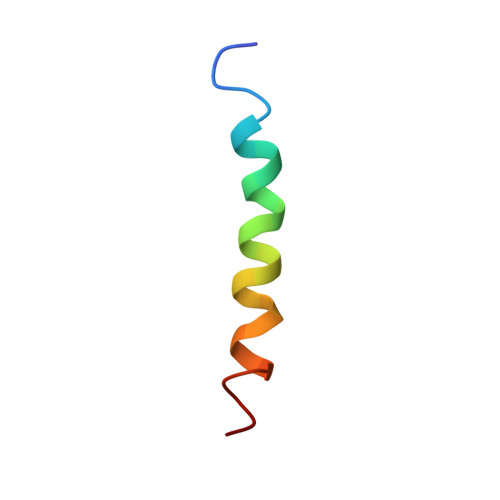
2AJJ, 2AJM, 2AJN, 2AJO - PubMed Abstract:
Hepatitis C virus (HCV) nonstructural protein 5A (NS5A) is a monotopic membrane protein anchored to the membrane by an N-terminal in-plane amphipathic alpha-helix. This membrane anchor is essential for the assembly of a functional viral replication complex. Although amino acid sequences differ considerably, putative membrane anchors with amphipathic features were predicted in NS5A from related Flaviviridae family members, in particular bovine viral diarrhea virus (BVDV), the prototype representative of the genus Pestivirus. We report here the NMR structure of the membrane anchor 1-28 of NS5A from BVDV in the presence of different membrane mimetic media. This anchor includes a long amphipathic alpha-helix of 21 residues interacting in-plane with the membrane interface and including a putative flexible region. Molecular dynamic simulation at a water-dodecane interface used to mimic the surface separating a lipid bilayer and an aqueous medium demonstrated the stability of the helix orientation and the location at the hydrophobic-hydrophilic interface. The flexible region of the helix appears to be required to allow the most favorable interaction of hydrophobic and hydrophilic side chain residues with their respective environment at the membrane interface. Despite the lack of amino acid sequence similarity, this amphipathic helix shares common structural features with that of the HCV counterpart, including a stable, hydrophobic N-terminal segment separated from the more hydrophilic C-terminal segment by a local, flexible region. These structural conservations point toward conserved roles of the N-terminal in-plane membrane anchors of NS5A in replication complex formation of HCV, BVDV, and other related viruses.
Organizational Affiliation:
Institut de Biologie et Chimie des Protéines, CNRS-UMR 5086, IFR128 BioSciences Lyon-Gerland, University of Lyon, France.