2AHR

Crystal Structures of 1-Pyrroline-5-Carboxylate Reductase from Human Pathogen Streptococcus pyogenes


Experimental Data Snapshot

  • Method: X-RAY DIFFRACTION
  • Resolution: 2.15 Å
  • R-Value Free: 0.210 
  • R-Value Work: 0.173 
  • R-Value Observed: 0.175 

wwPDB Validation   3D Report Full Report


Ligand Structure Quality Assessment 


This is version 1.3 of the entry. See complete history


Literature

Crystal Structures of Delta(1)-Pyrroline-5-carboxylate Reductase from Human Pathogens Neisseria meningitides and Streptococcus pyogenes

Nocek, B.Chang, C.Li, H.Lezondra, L.Holzle, D.Collart, F.Joachimiak, A.

(2005) J Mol Biol 354: 91-106

  • DOI: https://doi.org/10.1016/j.jmb.2005.08.036
  • Primary Citation of Related Structures:  
    1YQG, 2AG8, 2AHR, 2AMF

  • PubMed Abstract: 

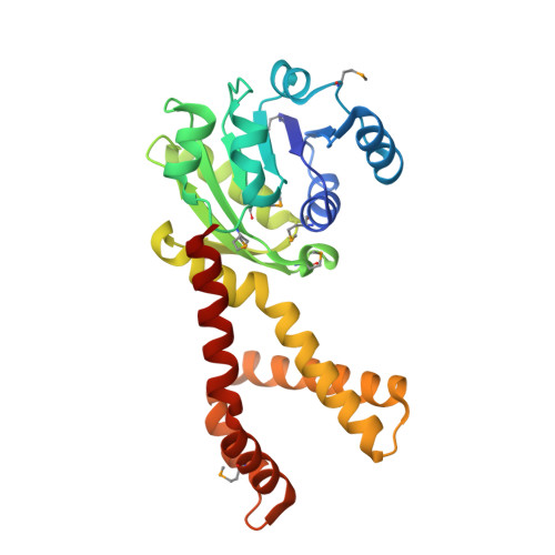
    L-proline is an amino acid that plays an important role in proteins uniquely contributing to protein folding, structure, and stability, and this amino acid serves as a sequence-recognition motif. Proline biosynthesis can occur via two pathways, one from glutamate and the other from arginine. In both pathways, the last step of biosynthesis, the conversion of delta1-pyrroline-5-carboxylate (P5C) to L-proline, is catalyzed by delta1-pyrroline-5-carboxylate reductase (P5CR) using NAD(P)H as a cofactor. We have determined the first crystal structure of P5CR from two human pathogens, Neisseria meningitides and Streptococcus pyogenes, at 2.0 angstroms and 2.15 angstroms resolution, respectively. The catalytic unit of P5CR is a dimer composed of two domains, but the biological unit seems to be species-specific. The N-terminal domain of P5CR is an alpha/beta/alpha sandwich, a Rossmann fold. The C-terminal dimerization domain is rich in alpha-helices and shows domain swapping. Comparison of the native structure of P5CR to structures complexed with L-proline and NADP+ in two quite different primary sequence backgrounds provides unique information about key functional features: the active site and the catalytic mechanism. The inhibitory L-proline has been observed in the crystal structure.


  • Organizational Affiliation

    Midwest Center for Structural Genomics and Structural Biology Center, Biosciences Division, Argonne National Laboratory, 9700 South Cass Avenue, Building 202, Argonne, IL 60439, USA.


Macromolecules
Find similar proteins by:  (by identity cutoff)  |  3D Structure
Entity ID: 1
MoleculeChains Sequence LengthOrganismDetailsImage
putative pyrroline carboxylate reductase
A, B, C, D, E
259Streptococcus pyogenes M1 GASMutation(s): 7 
EC: 1.5.1.2
UniProt
Find proteins for Q9A1S9 (Streptococcus pyogenes serotype M1)
Explore Q9A1S9 
Go to UniProtKB:  Q9A1S9
Entity Groups  
Sequence Clusters30% Identity50% Identity70% Identity90% Identity95% Identity100% Identity
UniProt GroupQ9A1S9
Sequence Annotations
Expand
  • Reference Sequence
Small Molecules
Ligands 3 Unique
IDChains Name / Formula / InChI Key2D Diagram3D Interactions
NAP
Query on NAP

Download Ideal Coordinates CCD File 
G [auth A],
J [auth B],
M [auth C],
P [auth D],
S [auth E]
NADP NICOTINAMIDE-ADENINE-DINUCLEOTIDE PHOSPHATE
C21 H28 N7 O17 P3
XJLXINKUBYWONI-NNYOXOHSSA-N
FMT
Query on FMT

Download Ideal Coordinates CCD File 
H [auth A],
K [auth B],
N [auth C],
Q [auth D],
T [auth E]
FORMIC ACID
C H2 O2
BDAGIHXWWSANSR-UHFFFAOYSA-N
NA
Query on NA

Download Ideal Coordinates CCD File 
F [auth A],
I [auth B],
L [auth C],
O [auth D],
R [auth E]
SODIUM ION
Na
FKNQFGJONOIPTF-UHFFFAOYSA-N
Modified Residues  1 Unique
IDChains TypeFormula2D DiagramParent
MSE
Query on MSE
A, B, C, D, E
L-PEPTIDE LINKINGC5 H11 N O2 SeMET
Experimental Data & Validation

Experimental Data

  • Method: X-RAY DIFFRACTION
  • Resolution: 2.15 Å
  • R-Value Free: 0.210 
  • R-Value Work: 0.173 
  • R-Value Observed: 0.175 
  • Space Group: C 1 2 1
Unit Cell:
Length ( Å )Angle ( ˚ )
a = 171.631α = 90
b = 109.646β = 96.08
c = 84.033γ = 90
Software Package:
Software NamePurpose
REFMACrefinement
SBC-Collectdata collection
HKL-3000data scaling
SHELXDphasing
SHELXEmodel building
SOLVEphasing
RESOLVEphasing
MLPHAREphasing
DMphasing
Omodel building
Cootmodel building
CCP4phasing

Structure Validation

View Full Validation Report


Ligand Structure Quality Assessment 


Entry History 

Deposition Data

Revision History  (Full details and data files)

  • Version 1.0: 2005-09-13
    Type: Initial release
  • Version 1.1: 2008-04-30
    Changes: Version format compliance
  • Version 1.2: 2011-07-13
    Changes: Advisory, Derived calculations, Source and taxonomy, Version format compliance
  • Version 1.3: 2017-10-11
    Changes: Refinement description