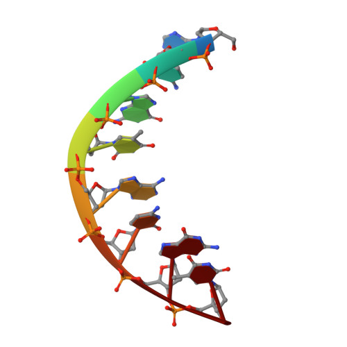
Structure of the DNA octanucleotide d(ACGTACGT)2.
Wilcock, D.J., Adams, A., Cardin, C.J., Wakelin, L.P.(1996) Acta Crystallogr D Biol Crystallogr 52: 481-485
- PubMed: 15299669
- DOI: https://doi.org/10.1107/S0907444995016052
- Primary Citation of Related Structures:
243D - PubMed Abstract:
d(ACGTACGT), C(78)H(84)N(30)O(32)P(7).20H(2)O, M(r) (DNA) = 2170, tetragonal, P4(3)2(1)2 (No 96), a = 42.845 (1), b = 42.845(1), c = 24.804 (1) A, V = 45532.5 (2) A(3), z = 8,lambda(MoKalpha) = 0.71069 A, micro (MoKalpha) = 0.10 mm(-1), T = 295 K, R = 0.18 for 1994 unique reflections between 5.0 and 1.9 A resolution. The self-complementary octanucleotide d(ACGTACGT)(2) has been crystallized and its structure determined to a resolution of 1.9 A. The asymmetric unit consists of a single strand of octamer with 20 water molecules. It is only the second example of an octanucleotide having terminal A.T base pairs whose structure has been determined by X-ray crystallography. The sequence adopts the modified A-type conformation found for all octanucleotide duplexes studied to date with the helix bent by approximately 15 degrees and an average tilt angle of 0 degrees. Unusually the data collection was carried out using a 3 kW molybdenum sealed-tube source. The conformational details are discussed in comparison with other closely related sequences.
Organizational Affiliation:
Department of Chemistry, Trinity College, Dublin, Ireland.