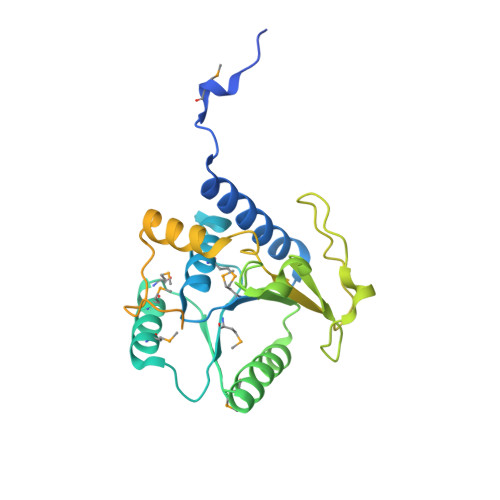
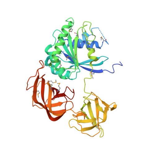
1ZUN

Crystal Structure of a GTP-Regulated ATP Sulfurylase Heterodimer from Pseudomonas syringae


Experimental Data Snapshot

  • Method: X-RAY DIFFRACTION
  • Resolution: 2.70 Å
  • R-Value Free: 0.276 
  • R-Value Work: 0.222 
  • R-Value Observed: 0.224 

wwPDB Validation   3D Report Full Report


Ligand Structure Quality Assessment 


This is version 1.2 of the entry. See complete history


Literature

Molecular basis for g protein control of the prokaryotic ATP sulfurylase.

Mougous, J.D.Lee, D.H.Hubbard, S.C.Schelle, M.W.Vocadlo, D.J.Berger, J.M.Bertozzi, C.R.

(2006) Mol Cell 21: 109-122

  • DOI: https://doi.org/10.1016/j.molcel.2005.10.034
  • Primary Citation of Related Structures:  
    1ZUN

  • PubMed Abstract: 

    Sulfate assimilation is a critical component of both primary and secondary metabolism. An essential step in this pathway is the activation of sulfate through adenylation by the enzyme ATP sulfurylase (ATPS), forming adenosine 5'-phosphosulfate (APS). Proteobacterial ATPS overcomes this energetically unfavorable reaction by associating with a regulatory G protein, coupling the energy of GTP hydrolysis to APS formation. To discover the molecular basis of this unusual role for a G protein, we biochemically characterized and solved the X-ray crystal structure of a complex between Pseudomonas syringae ATPS (CysD) and its associated regulatory G protein (CysN). The structure of CysN*D shows the two proteins in tight association; however, the nucleotides bound to each subunit are spatially segregated. We provide evidence that conserved switch motifs in the G domain of CysN allosterically mediate interactions between the nucleotide binding sites. This structure suggests a molecular mechanism by which conserved G domain architecture is used to energetically link GTP turnover to the production of an essential metabolite.


  • Organizational Affiliation

    Howard Hughes Medical Institute, University of California, Berkeley, Berkeley, California 94720, USA.


Macromolecules
Find similar proteins by:  (by identity cutoff)  |  3D Structure
Entity ID: 1
MoleculeChains Sequence LengthOrganismDetailsImage
Sulfate adenylyltransferase subunit 2325Pseudomonas syringaeMutation(s): 8 
Gene Names: cysD
EC: 2.7.7.4
UniProt
Find proteins for Q87WW0 (Pseudomonas syringae pv. tomato (strain ATCC BAA-871 / DC3000))
Explore Q87WW0 
Go to UniProtKB:  Q87WW0
Entity Groups  
Sequence Clusters30% Identity50% Identity70% Identity90% Identity95% Identity100% Identity
UniProt GroupQ87WW0
Sequence Annotations
Expand
  • Reference Sequence
Find similar proteins by:  (by identity cutoff)  |  3D Structure
Entity ID: 2
MoleculeChains Sequence LengthOrganismDetailsImage
sulfate adenylate transferase, subunit 1/adenylylsulfate kinase434Pseudomonas syringae pv. tomato str. DC3000Mutation(s): 12 
Gene Names: cysNC
UniProt
Find proteins for Q87WW1 (Pseudomonas syringae pv. tomato (strain ATCC BAA-871 / DC3000))
Explore Q87WW1 
Go to UniProtKB:  Q87WW1
Entity Groups  
Sequence Clusters30% Identity50% Identity70% Identity90% Identity95% Identity100% Identity
UniProt GroupQ87WW1
Sequence Annotations
Expand
  • Reference Sequence
Small Molecules
Modified Residues  1 Unique
IDChains TypeFormula2D DiagramParent
MSE
Query on MSE
A
L-PEPTIDE LINKINGC5 H11 N O2 SeMET
Experimental Data & Validation

Experimental Data

  • Method: X-RAY DIFFRACTION
  • Resolution: 2.70 Å
  • R-Value Free: 0.276 
  • R-Value Work: 0.222 
  • R-Value Observed: 0.224 
  • Space Group: H 3
Unit Cell:
Length ( Å )Angle ( ˚ )
a = 110.072α = 90
b = 110.072β = 90
c = 171.002γ = 120
Software Package:
Software NamePurpose
REFMACrefinement
HKL-2000data reduction
SCALEPACKdata scaling
SOLVEphasing

Structure Validation

View Full Validation Report


Ligand Structure Quality Assessment 


Entry History 

Deposition Data

Revision History  (Full details and data files)

  • Version 1.0: 2006-01-17
    Type: Initial release
  • Version 1.1: 2008-04-30
    Changes: Version format compliance
  • Version 1.2: 2011-07-13
    Changes: Version format compliance