The role of the divalent cation in the structure of the I domain from the CD11a/CD18 integrin.
Qu, A., Leahy, D.J.(1996) Structure 4: 931-942
- PubMed: 8805579
- DOI: https://doi.org/10.1016/s0969-2126(96)00100-1
- Primary Citation of Related Structures:
1ZON, 1ZOO, 1ZOP - PubMed Abstract:
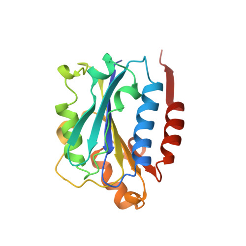
The integrin family of cell-surface receptors mediates a wide variety of cell-cell and cell-extracellular matrix interactions. Integrin-ligand interactions are invariably dependent on the presence of divalent cations, and a subset of integrins contain a approximately 200 amino acid inserted (I) domain that is important for ligand binding activity and contains a single divalent cation binding site. Many integrins are believed to respond to stimuli by undergoing a conformational change that increases their affinity for ligand, and there is a clear difference between two crystal structures of the CD11b I domain with different divalent cations (magnesium and manganese) bound. In addition to the different bound cation, a 'ligand mimetic' crystal lattice interaction in the CD11b I domain structure with bound magnesium has led to the interpretation that the different CD11b I domain structures represent different affinity states of I domains. The influence of the bound cation on I domain structure and function remains incompletely understood, however. The crystal structure of the CD11a I domain bound to manganese is known. We therefore set out to determine whether this structure changes when the metal ion is altered or removed. We report here the crystal structures of the CD11a I domain determined in the absence of bound metal ion and with bound magnesium ion. No major structural rearrangements are observed in the metal-binding site of the CD11a I domain in the absence or presence of bound manganese ion. The structures of the CD11a I domain with magnesium or manganese bound are extremely similar. The conformation of the CD11a I domain is not altered by changes in metal ion binding. The cation-dependence of ligand binding thus indicates that the metal ion is either involved in direct interaction with ligand or required to promote a favorable quaternary arrangement of the integrin.
Organizational Affiliation:
Department of Biophysics and Biophysical Chemistry, Johns Hopkins University School of Medicine, 725 N. Wolfe Street, Baltimore, MD 21205, USA.