Structure of the SH3 domain of human osteoclast-stimulating factor at atomic resolution.
Chen, L., Wang, Y., Wells, D., Toh, D., Harold, H., Zhou, J., DiGiammarino, E., Meehan, E.J.(2006) Acta Crystallogr Sect F Struct Biol Cryst Commun 62: 844-848
- PubMed: 16946461
- DOI: https://doi.org/10.1107/S1744309106030004
- Primary Citation of Related Structures:
1ZLM - PubMed Abstract:
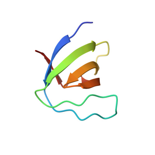
Osteoclast-stimulating factor (OSF) is an intracellular signaling protein, produced by osteoclasts themselves, that enhances osteoclast formation and bone resorption. It is thought to act via an Src-related signaling pathway and contains SH3 and ankyrin-repeat domains which are involved in protein-protein interactions. As part of a structure-based anti-bone-loss drug-design program, the atomic resolution X-ray structure of the recombinant human OSF SH3 domain (hOSF-SH3) has been determined. The domain, residues 12-72, yielded crystals that diffracted to the ultrahigh resolution of 1.07 A. The overall structure shows a characteristic SH3 fold consisting of two perpendicular beta-sheets that form a beta-barrel. Structure-based sequence alignment reveals that the putative proline-rich peptide-binding site of hOSF-SH3 consists of (i) residues that are highly conserved in the SH3-domain family, including residues Tyr21, Phe23, Trp49, Pro62, Asn64 and Tyr65, and (ii) residues that are less conserved and/or even specific to hOSF, including Thr22, Arg26, Thr27, Glu30, Asp46, Thr47, Asn48 and Leu60, which might be key to designing specific inhibitors for hOSF to fight osteoporosis and related bone-loss diseases. There are a total of 13 well defined water molecules forming hydrogen bonds with the above residues in and around the peptide-binding pocket. Some of those water molecules might be important for drug-design approaches. The hOSF-SH3 structure at atomic resolution provides an accurate framework for structure-based design of its inhibitors.
Organizational Affiliation:
Laboratory for Structural Biology, University of Alabama in Huntsville, Huntsville, Alabama 35899, USA. chenlq@uah.edu