1Z5Y

Crystal Structure Of The Disulfide-Linked Complex Between The N-Terminal Domain Of The Electron Transfer Catalyst DsbD and The Cytochrome c Biogenesis Protein CcmG


Experimental Data Snapshot

  • Method: X-RAY DIFFRACTION
  • Resolution: 1.94 Å
  • R-Value Free: 0.270 
  • R-Value Work: 0.235 

wwPDB Validation   3D Report Full Report


This is version 1.4 of the entry. See complete history


Literature

Structural Basis and Kinetics of DsbD-Dependent Cytochrome c Maturation

Stirnimann, C.U.Rozhkova, A.Grauschopf, U.Gruetter, M.G.Glockshuber, R.Capitani, G.

(2005) Structure 13: 985-993

  • DOI: https://doi.org/10.1016/j.str.2005.04.014
  • Primary Citation of Related Structures:  
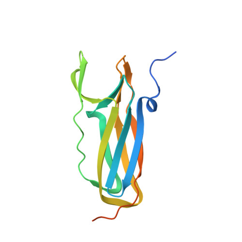
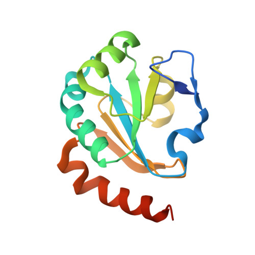
    1Z5Y

  • PubMed Abstract: 

    DsbD from Escherichia coli transports two electrons from cytoplasmic thioredoxin to the periplasmic substrate proteins DsbC, DsbG and CcmG. DsbD consists of an N-terminal periplasmic domain (nDsbD), a C-terminal periplasmic domain, and a central transmembrane domain. Each domain possesses two cysteines required for electron transport. Herein, we demonstrate fast (3.9 x 10(5) M(-1)s(-1)) and direct disulfide exchange between nDsbD and CcmG, a highly specific disulfide reductase essential for cytochrome c maturation. We determined the crystal structure of the disulfide-linked complex between nDsbD and the soluble part of CcmG at 1.94 A resolution. In contrast to the other two known complexes of nDsbD with target proteins, the N-terminal segment of nDsbD contributes to specific recognition of CcmG. This and other features, like the possibility of using an additional interaction surface, constitute the structural basis for the adaptability of nDsbD to different protein substrates.


  • Organizational Affiliation

    Biochemisches Institut, Universität Zürich, Winterthurerstrasse 190, 8057 Zürich, Switzerland.


Macromolecules
Find similar proteins by:  (by identity cutoff)  |  3D Structure
Entity ID: 1
MoleculeChains Sequence LengthOrganismDetailsImage
Thiol:disulfide interchange protein dsbDA [auth D]143Escherichia coliMutation(s): 1 
Gene Names: DSBDDIPZCYCZCUTA2B4136
EC: 1.8.1.8
UniProt
Find proteins for P36655 (Escherichia coli (strain K12))
Explore P36655 
Go to UniProtKB:  P36655
Entity Groups  
Sequence Clusters30% Identity50% Identity70% Identity90% Identity95% Identity100% Identity
UniProt GroupP36655
Sequence Annotations
Expand
  • Reference Sequence
Find similar proteins by:  (by identity cutoff)  |  3D Structure
Entity ID: 2
MoleculeChains Sequence LengthOrganismDetailsImage
Thiol:disulfide interchange protein dsbEB [auth E]149Escherichia coliMutation(s): 1 
Gene Names: DSBECCMG
UniProt
Find proteins for P0AA86 (Escherichia coli (strain K12))
Explore P0AA86 
Go to UniProtKB:  P0AA86
Entity Groups  
Sequence Clusters30% Identity50% Identity70% Identity90% Identity95% Identity100% Identity
UniProt GroupP0AA86
Sequence Annotations
Expand
  • Reference Sequence
Experimental Data & Validation

Experimental Data

  • Method: X-RAY DIFFRACTION
  • Resolution: 1.94 Å
  • R-Value Free: 0.270 
  • R-Value Work: 0.235 
  • Space Group: P 1 21 1
Unit Cell:
Length ( Å )Angle ( ˚ )
a = 53.574α = 90
b = 53.165β = 104.88
c = 63.833γ = 90
Software Package:
Software NamePurpose
DENZOdata reduction
SCALEPACKdata scaling
PHASERphasing
CNSrefinement

Structure Validation

View Full Validation Report


Entry History 

Deposition Data

Revision History  (Full details and data files)

  • Version 1.0: 2005-07-19
    Type: Initial release
  • Version 1.1: 2008-04-30
    Changes: Version format compliance
  • Version 1.2: 2011-07-13
    Changes: Version format compliance
  • Version 1.3: 2021-11-10
    Changes: Database references, Derived calculations
  • Version 1.4: 2023-10-25
    Changes: Data collection, Refinement description