1YV3

The structural basis of blebbistatin inhibition and specificity for myosin II


Experimental Data Snapshot

  • Method: X-RAY DIFFRACTION
  • Resolution: 2.00 Å
  • R-Value Free: 0.215 
  • R-Value Work: 0.173 
  • R-Value Observed: 0.175 

wwPDB Validation   3D Report Full Report


Ligand Structure Quality Assessment 


This is version 1.3 of the entry. See complete history


Literature

The structural basis of blebbistatin inhibition and specificity for myosin II.

Allingham, J.S.Smith, R.Rayment, I.

(2005) Nat Struct Mol Biol 12: 378-379

  • DOI: https://doi.org/10.1038/nsmb908
  • Primary Citation of Related Structures:  
    1YV3

  • PubMed Abstract: 

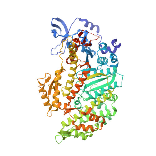
    Molecular motors play a central role in cytoskeletal-mediated cellular processes and thus present an excellent target for cellular control by pharmacological agents. Yet very few such compounds have been found. We report here the structure of blebbistatin, which inhibits specific myosin isoforms, bound to the motor domain of Dictyostelium discoideum myosin II. This reveals the structural basis for its specificity and provides insight into the development of new agents.


  • Organizational Affiliation

    Department of Biochemistry, University of Wisconsin, 433 Babcock Drive, Madison, Wisconsin 53706, USA.


Macromolecules
Find similar proteins by:  (by identity cutoff)  |  3D Structure
Entity ID: 1
MoleculeChains Sequence LengthOrganismDetailsImage
Myosin II heavy chain762Dictyostelium discoideumMutation(s): 0 
UniProt
Find proteins for P08799 (Dictyostelium discoideum)
Explore P08799 
Go to UniProtKB:  P08799
Entity Groups  
Sequence Clusters30% Identity50% Identity70% Identity90% Identity95% Identity100% Identity
UniProt GroupP08799
Sequence Annotations
Expand
  • Reference Sequence
Small Molecules
Ligands 5 Unique
IDChains Name / Formula / InChI Key2D Diagram3D Interactions
ADP
Query on ADP

Download Ideal Coordinates CCD File 
E [auth A]ADENOSINE-5'-DIPHOSPHATE
C10 H15 N5 O10 P2
XTWYTFMLZFPYCI-KQYNXXCUSA-N
BIT
Query on BIT

Download Ideal Coordinates CCD File 
F [auth A](-)-1-PHENYL-1,2,3,4-TETRAHYDRO-4-HYDROXYPYRROLO[2,3-B]-7-METHYLQUINOLIN-4-ONE
C18 H16 N2 O2
LZAXPYOBKSJSEX-GOSISDBHSA-N
VO4
Query on VO4

Download Ideal Coordinates CCD File 
C [auth A]VANADATE ION
O4 V
LSGOVYNHVSXFFJ-UHFFFAOYSA-N
EDO
Query on EDO

Download Ideal Coordinates CCD File 
G [auth A]1,2-ETHANEDIOL
C2 H6 O2
LYCAIKOWRPUZTN-UHFFFAOYSA-N
MG
Query on MG

Download Ideal Coordinates CCD File 
B [auth A],
D [auth A]
MAGNESIUM ION
Mg
JLVVSXFLKOJNIY-UHFFFAOYSA-N
Binding Affinity Annotations 
IDSourceBinding Affinity
BIT BindingDB:  1YV3 IC50: 4900 (nM) from 1 assay(s)
Experimental Data & Validation

Experimental Data

  • Method: X-RAY DIFFRACTION
  • Resolution: 2.00 Å
  • R-Value Free: 0.215 
  • R-Value Work: 0.173 
  • R-Value Observed: 0.175 
  • Space Group: C 2 2 21
Unit Cell:
Length ( Å )Angle ( ˚ )
a = 88.106α = 90
b = 145.77β = 90
c = 152.912γ = 90
Software Package:
Software NamePurpose
REFMACrefinement
HKL-2000data reduction
SCALEPACKdata scaling
MOLREPphasing

Structure Validation

View Full Validation Report


Ligand Structure Quality Assessment 


Entry History 

Deposition Data

Revision History  (Full details and data files)

  • Version 1.0: 2005-03-08
    Type: Initial release
  • Version 1.1: 2008-04-30
    Changes: Version format compliance
  • Version 1.2: 2011-07-13
    Changes: Version format compliance
  • Version 1.3: 2023-08-23
    Changes: Data collection, Database references, Derived calculations, Refinement description, Structure summary