1YU7

Crystal Structure of the W64Y mutant of Villin Headpiece


Experimental Data Snapshot

  • Method: X-RAY DIFFRACTION
  • Resolution: 1.50 Å
  • R-Value Free: 0.210 
  • R-Value Work: 0.192 
  • R-Value Observed: 0.194 

wwPDB Validation   3D Report Full Report


This is version 1.5 of the entry. See complete history


Literature

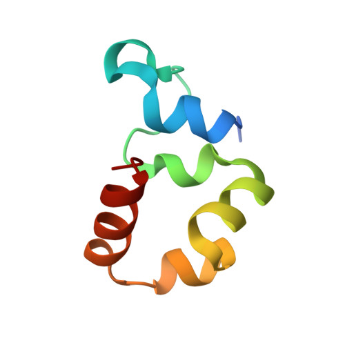
High-resolution crystal structures of villin headpiece and mutants with reduced F-actin binding activity.

Meng, J.Vardar, D.Wang, Y.Guo, H.C.Head, J.F.McKnight, C.J.

(2005) Biochemistry 44: 11963-11973

  • DOI: https://doi.org/10.1021/bi050850x
  • Primary Citation of Related Structures:  
    1YU5, 1YU7, 1YU8

  • PubMed Abstract: 

    Villin-type headpiece domains are approximately 70 amino acid modular motifs found at the C terminus of a variety of actin cytoskeleton-associated proteins. The headpiece domain of villin, a protein found in the actin bundles of the brush border epithelium, is of interest both as a compact F-actin binding domain and as a model folded protein. We have determined the high-resolution crystal structures of chicken villin headpiece (HP67) at 1.4 A resolution as well as two mutants, R37A and W64Y, at 1.45 and 1.5 A resolution, respectively. Replacement of R37 causes a 5-fold reduction in F-actin binding affinity in sedimentation assays. Replacement of W64 results in a much more drastic reduction in F-actin binding affinity without significant changes in headpiece structure or stability. The detailed comparison of these crystal structures with each other and to our previously determined NMR structures of HP67 and the 35-residue autonomously folding subdomain in villin headpiece, HP35, provides the details of the headpiece fold and further defines the F-actin binding site of villin-type headpiece domains.


  • Organizational Affiliation

    Department of Physiology and Biophysics, Boston University School of Medicine, 715 Albany Street, Boston, Massachusetts 02118, USA.


Macromolecules
Find similar proteins by:  (by identity cutoff)  |  3D Structure
Entity ID: 1
MoleculeChains Sequence LengthOrganismDetailsImage
VillinA [auth X]67Gallus gallusMutation(s): 1 
UniProt
Find proteins for P02640 (Gallus gallus)
Explore P02640 
Go to UniProtKB:  P02640
Entity Groups  
Sequence Clusters30% Identity50% Identity70% Identity90% Identity95% Identity100% Identity
UniProt GroupP02640
Sequence Annotations
Expand
  • Reference Sequence
Experimental Data & Validation

Experimental Data

  • Method: X-RAY DIFFRACTION
  • Resolution: 1.50 Å
  • R-Value Free: 0.210 
  • R-Value Work: 0.192 
  • R-Value Observed: 0.194 
  • Space Group: P 21 21 21
Unit Cell:
Length ( Å )Angle ( ˚ )
a = 31.419α = 90
b = 38.259β = 90
c = 52.807γ = 90
Software Package:
Software NamePurpose
HKL-2000data collection
SCALEPACKdata scaling
CNSrefinement
HKL-2000data reduction
CNSphasing

Structure Validation

View Full Validation Report


Entry History 

Deposition Data

Revision History  (Full details and data files)

  • Version 1.0: 2005-09-06
    Type: Initial release
  • Version 1.1: 2008-04-30
    Changes: Version format compliance
  • Version 1.2: 2011-07-13
    Changes: Version format compliance
  • Version 1.3: 2021-10-20
    Changes: Database references
  • Version 1.4: 2024-02-14
    Changes: Data collection
  • Version 1.5: 2024-04-03
    Changes: Refinement description