1YMH

anti-HCV Fab 19D9D6 complexed with protein L (PpL) mutant A66W


Experimental Data Snapshot

  • Method: X-RAY DIFFRACTION
  • Resolution: 2.60 Å
  • R-Value Free: 0.302 
  • R-Value Work: 0.224 
  • R-Value Observed: 0.228 

wwPDB Validation   3D Report Full Report


This is version 1.3 of the entry. See complete history


Literature

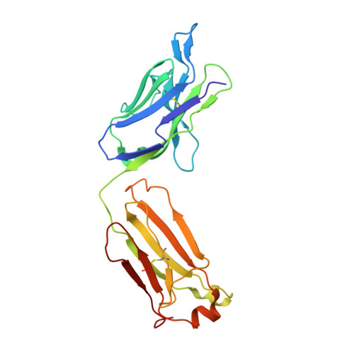
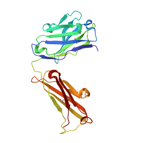
Comparison of the crystallization and crystal packing of two Fab single-site mutant protein L complexes.

Granata, V.Housden, N.G.Harrison, S.Jolivet-Reynaud, C.Gore, M.G.Stura, E.A.

(2005) Acta Crystallogr D Biol Crystallogr 61: 750-754

  • DOI: https://doi.org/10.1107/S0907444905007110
  • Primary Citation of Related Structures:  
    1YMH

  • PubMed Abstract: 

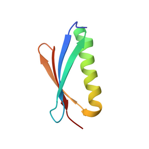
    Protein L from Peptostreptococcus magnus (PpL) is a multidomain protein composed of four or five immunoglobulin-binding domains that target the kappa light chain of a large repertoire of human and murine antibodies. Thus, a single domain of this protein can be used to aid the crystallization of Fab, free or complexed to their antigen when it is not possible to obtain crystals without it. Each wild-type PpL domain has two light-chain binding sites that target the same region of the light chain and can thus bring together two Fab-antigen complexes within the crystal lattice. In this context the small PpL domain is sandwiched between two Fab and cannot participate in crystal contacts, thus mutants are unlikely to increase the chances of crystallizing a particular complex. However, it is possible to design mutants that can bind at only one site by making use of the crystal structures obtained so far. Such mutants will have a free surface that can participate in crystal contacts and that can be modified to improve its crystal contact-forming properties. Here, a comparison of two single-site mutants that differ at three different positions is reported. In both mutants two different tryptophan residues participate in crystal-packing interactions, suggesting that this residue may be particularly interesting for enhancing crystal-contact formation.


  • Organizational Affiliation

    Università Federico II, Dipartimento di Chimica Biologica, Via Mezzocannone, Napoli, Italy.


Macromolecules
Find similar proteins by:  (by identity cutoff)  |  3D Structure
Entity ID: 1
MoleculeChains Sequence LengthOrganismDetailsImage
Fab 16D9D6, light chainA,
D [auth C]
220Mus musculusMutation(s): 0 
Entity Groups  
Sequence Clusters30% Identity50% Identity70% Identity90% Identity95% Identity100% Identity
Sequence Annotations
Expand
  • Reference Sequence
Find similar proteins by:  (by identity cutoff)  |  3D Structure
Entity ID: 2
MoleculeChains Sequence LengthOrganismDetailsImage
Fab 16D9D6, heavy chainB,
E [auth D]
218Mus musculusMutation(s): 0 
Entity Groups  
Sequence Clusters30% Identity50% Identity70% Identity90% Identity95% Identity100% Identity
Sequence Annotations
Expand
  • Reference Sequence
Find similar proteins by:  (by identity cutoff)  |  3D Structure
Entity ID: 3
MoleculeChains Sequence LengthOrganismDetailsImage
Protein LC [auth E],
F
65Finegoldia magna ATCC 29328Mutation(s): 0 
UniProt
Find proteins for Q51918 (Finegoldia magna)
Explore Q51918 
Go to UniProtKB:  Q51918
Entity Groups  
Sequence Clusters30% Identity50% Identity70% Identity90% Identity95% Identity100% Identity
UniProt GroupQ51918
Sequence Annotations
Expand
  • Reference Sequence
Experimental Data & Validation

Experimental Data

  • Method: X-RAY DIFFRACTION
  • Resolution: 2.60 Å
  • R-Value Free: 0.302 
  • R-Value Work: 0.224 
  • R-Value Observed: 0.228 
  • Space Group: P 21 21 21
Unit Cell:
Length ( Å )Angle ( ˚ )
a = 77.143α = 90
b = 111.469β = 90
c = 148.698γ = 90
Software Package:
Software NamePurpose
REFMACrefinement
DENZOdata reduction
SCALEPACKdata scaling
MOLREPphasing

Structure Validation

View Full Validation Report


Entry History 

Deposition Data

Revision History  (Full details and data files)

  • Version 1.0: 2005-05-31
    Type: Initial release
  • Version 1.1: 2008-04-30
    Changes: Version format compliance
  • Version 1.2: 2011-07-13
    Changes: Source and taxonomy, Version format compliance
  • Version 1.3: 2023-10-25
    Changes: Data collection, Database references, Refinement description