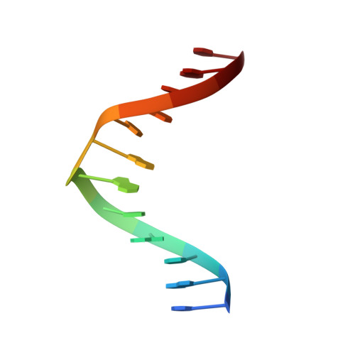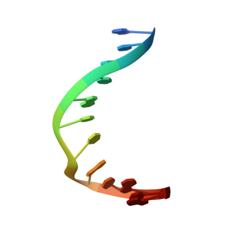
NMR solution structures of bistranded abasic site lesions in DNA.
Hazel, R.D., Tian, K., de Los Santos, C.(2008) Biochemistry 47: 11909-11919
- PubMed: 18950195
- DOI: https://doi.org/10.1021/bi800950t
- Primary Citation of Related Structures:
1YCT, 1YCW - PubMed Abstract:
Ionizing radiation produces clustered lesions in DNA. Since the orientation of bistranded lesions affects their recognition by DNA repair enzymes, clustered damages are more difficult to process and thus more toxic than single oxidative lesions. In order to understand the structural determinants that lead to differential recognition, we used NMR spectroscopy and restrained molecular dynamics to solve the structure of two DNA duplexes, each containing two stable abasic site analogues positioned on opposite strands of the duplex and staggered in the 3' (-1 duplex, (AP) 2-1 duplex) or 5' (+1 duplex, (AP) 2+1 duplex) direction. Cross-peak connectivities observed in the nonexchangeable NOESY spectra indicate compression of the helix at the lesion site of the duplexes, resulting in the formation of two abasic bulges. The exchangeable proton spectra show the AP site partner nucleotides forming interstrand hydrogen bonds that are characteristic of a Watson-Crick G.C base pairs, confirming the extra helical nature of the AP residues. Restrained molecular dynamics simulations generate a set of converging structures in full agreement with the spectroscopic data. In the (AP) 2-1 duplex, the extra helical abasic site residues reside in the minor groove of the helix, while they appear in the major groove in the (AP) 2+1 duplex. These structural differences are consistent with the differential recognition of bistranded abasic site lesions by human AP endonuclease.
Organizational Affiliation:
Department of Physiology & Biophysics, and Department of Pharmacological Sciences, Stony Brook University, School of Medicine, Stony Brook, New York 11794-8651, USA.