1Y56

Crystal structure of L-proline dehydrogenase from P.horikoshii


Experimental Data Snapshot

  • Method: X-RAY DIFFRACTION
  • Resolution: 2.86 Å
  • R-Value Free: 0.226 
  • R-Value Work: 0.182 
  • R-Value Observed: 0.184 

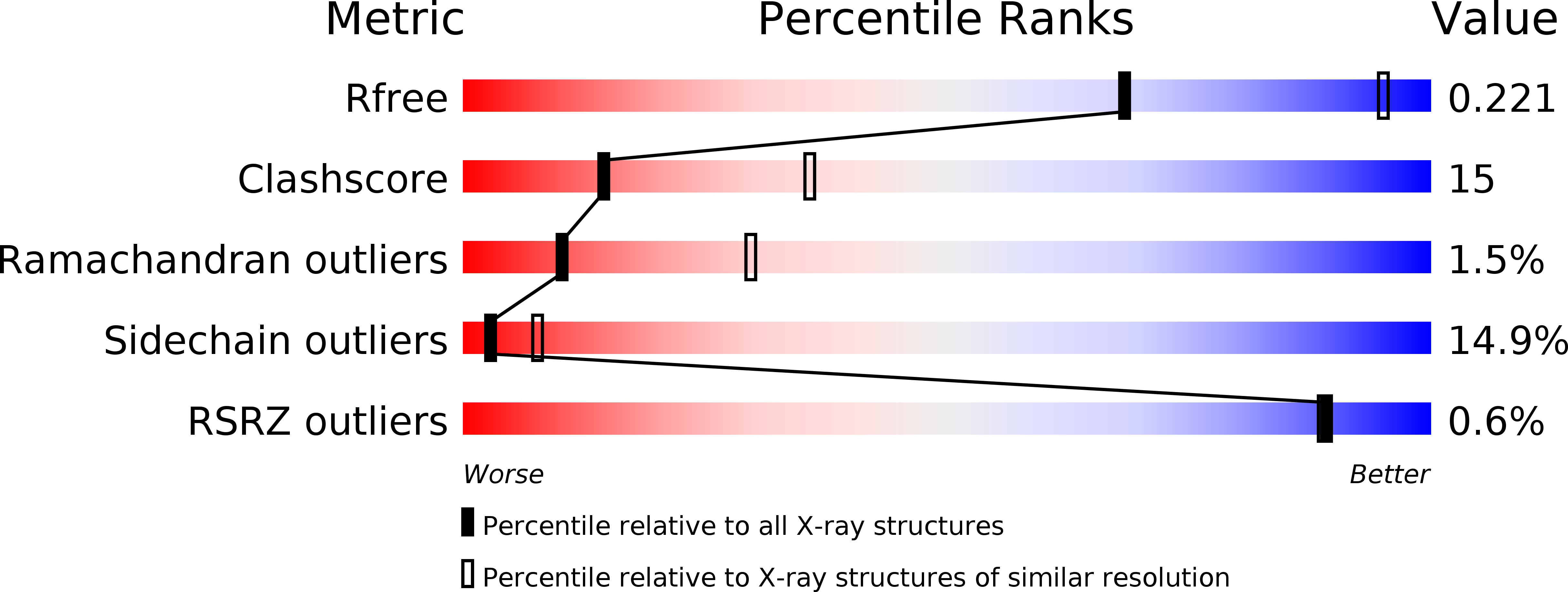
wwPDB Validation   3D Report Full Report


Ligand Structure Quality Assessment 


This is version 1.2 of the entry. See complete history


Literature

Crystal structure of a novel FAD-, FMN-, and ATP-containing L-proline dehydrogenase complex from Pyrococcus horikoshii

Tsuge, H.Kawakami, R.Sakuraba, H.Ago, H.Miyano, M.Aki, K.Katunuma, N.Ohshima, T.

(2005) J Biol Chem 280: 31045-31049

  • DOI: https://doi.org/10.1074/jbc.C500234200
  • Primary Citation of Related Structures:  
    1Y56

  • PubMed Abstract: 

    Two novel types of dye-linked L-proline dehydrogenase complex (PDH1 and PDH2) were found in a hyperthermophilic archaeon, Pyrococcus horikoshii OT3. Here we report the first crystal structure of PDH1, which is a heterooctameric complex (alphabeta)4 containing three different cofactors: FAD, FMN, and ATP. The structure was determined by x-ray crystallography to a resolution of 2.86 angstroms. The structure of the beta subunit, which is an L-proline dehydrogenase catalytic component containing FAD as a cofactor, was similar to that of monomeric sarcosine oxidase. On the other hand, the alpha subunit possessed a unique structure composed of a classical dinucleotide fold domain with ATP, a central domain, an N-terminal domain, and a Cys-clustered domain. Serving as a third cofactor, FMN was located at the interface between the alpha and beta subunits in a novel configuration. The observed structure suggests that FAD and FMN are incorporated into an electron transfer system, with electrons passing from the former to the latter. The function of ATP is unknown, but it may play a regulatory role. Although the structure of the alpha subunit differs from that of the beta subunit, except for the presence of an analogous dinucleotide domain with a different cofactor, the structural characteristics of PDH1 suggest that each represents a divergent enzyme that arose from a common ancestral flavoenzyme and that they eventually formed a complex to gain a new function. The structural characteristics described here reveal the PDH1 complex to be a unique diflavin dehydrogenase containing a novel electron transfer system.


  • Organizational Affiliation

    Institute for Health Sciences, Tokushima Bunri University, 180 Nishihama-bouji, Yamashiro-cho, Tokushima 770-8514, Japan. tsuge@tokushima.bunri-u.ac.jp


Macromolecules
Find similar proteins by:  (by identity cutoff)  |  3D Structure
Entity ID: 1
MoleculeChains Sequence LengthOrganismDetailsImage
hypothetical protein PH1363493Pyrococcus horikoshii OT3Mutation(s): 0 
EC: 1.5.99.8
UniProt
Find proteins for O59088 (Pyrococcus horikoshii (strain ATCC 700860 / DSM 12428 / JCM 9974 / NBRC 100139 / OT-3))
Explore O59088 
Go to UniProtKB:  O59088
Entity Groups  
Sequence Clusters30% Identity50% Identity70% Identity90% Identity95% Identity100% Identity
UniProt GroupO59088
Sequence Annotations
Expand
  • Reference Sequence
Find similar proteins by:  (by identity cutoff)  |  3D Structure
Entity ID: 2
MoleculeChains Sequence LengthOrganismDetailsImage
sarcosine oxidase382Pyrococcus horikoshii OT3Mutation(s): 0 
EC: 1.5.99.8
UniProt
Find proteins for O59089 (Pyrococcus horikoshii (strain ATCC 700860 / DSM 12428 / JCM 9974 / NBRC 100139 / OT-3))
Explore O59089 
Go to UniProtKB:  O59089
Entity Groups  
Sequence Clusters30% Identity50% Identity70% Identity90% Identity95% Identity100% Identity
UniProt GroupO59089
Sequence Annotations
Expand
  • Reference Sequence
Small Molecules
Ligands 7 Unique
IDChains Name / Formula / InChI Key2D Diagram3D Interactions
FAD
Query on FAD

Download Ideal Coordinates CCD File 
I [auth B]FLAVIN-ADENINE DINUCLEOTIDE
C27 H33 N9 O15 P2
VWWQXMAJTJZDQX-UYBVJOGSSA-N
ATP
Query on ATP

Download Ideal Coordinates CCD File 
F [auth A]ADENOSINE-5'-TRIPHOSPHATE
C10 H16 N5 O13 P3
ZKHQWZAMYRWXGA-KQYNXXCUSA-N
FMN
Query on FMN

Download Ideal Coordinates CCD File 
E [auth A]FLAVIN MONONUCLEOTIDE
C17 H21 N4 O9 P
FVTCRASFADXXNN-SCRDCRAPSA-N
CXS
Query on CXS

Download Ideal Coordinates CCD File 
G [auth A]3-CYCLOHEXYL-1-PROPYLSULFONIC ACID
C9 H19 N O3 S
PJWWRFATQTVXHA-UHFFFAOYSA-N
SO4
Query on SO4

Download Ideal Coordinates CCD File 
D [auth A]SULFATE ION
O4 S
QAOWNCQODCNURD-UHFFFAOYSA-L
FE
Query on FE

Download Ideal Coordinates CCD File 
C [auth A]FE (III) ION
Fe
VTLYFUHAOXGGBS-UHFFFAOYSA-N
CL
Query on CL

Download Ideal Coordinates CCD File 
H [auth B]CHLORIDE ION
Cl
VEXZGXHMUGYJMC-UHFFFAOYSA-M
Experimental Data & Validation

Experimental Data

  • Method: X-RAY DIFFRACTION
  • Resolution: 2.86 Å
  • R-Value Free: 0.226 
  • R-Value Work: 0.182 
  • R-Value Observed: 0.184 
  • Space Group: P 64 2 2
Unit Cell:
Length ( Å )Angle ( ˚ )
a = 172.219α = 90
b = 172.219β = 90
c = 175.398γ = 120
Software Package:
Software NamePurpose
REFMACrefinement
HKL-2000data reduction
SCALEPACKdata scaling
SOLVEphasing

Structure Validation

View Full Validation Report


Ligand Structure Quality Assessment 


Entry History 

Deposition Data

Revision History  (Full details and data files)

  • Version 1.0: 2005-07-26
    Type: Initial release
  • Version 1.1: 2008-04-30
    Changes: Version format compliance
  • Version 1.2: 2011-07-13
    Changes: Derived calculations, Source and taxonomy, Version format compliance