1Y2O

Structure of N-terminal domain IRSp53/BAIAP2


Experimental Data Snapshot

  • Method: X-RAY DIFFRACTION
  • Resolution: 2.20 Å
  • R-Value Free: 0.267 
  • R-Value Work: 0.228 
  • R-Value Observed: 0.230 

wwPDB Validation   3D Report Full Report


This is version 1.2 of the entry. See complete history


Literature

Structural basis of filopodia formation induced by the IRSp53/MIM homology domain of human IRSp53

Millard, T.H.Bompard, G.Heung, M.-Y.Dafforn, T.R.Scott, D.J.Machesky, L.M.Futterer, K.

(2005) EMBO J 24: 240-250

  • DOI: https://doi.org/10.1038/sj.emboj.7600535
  • Primary Citation of Related Structures:  
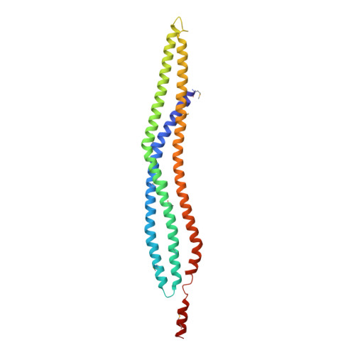
    1Y2O

  • PubMed Abstract: 

    The scaffolding protein insulin receptor tyrosine kinase substrate p53 (IRSp53), a ubiquitous regulator of the actin cytoskeleton, mediates filopodia formation under the control of Rho-family GTPases. IRSp53 comprises a central SH3 domain, which binds to proline-rich regions of a wide range of actin regulators, and a conserved N-terminal IRSp53/MIM homology domain (IMD) that harbours F-actin-bundling activity. Here, we present the crystal structure of this novel actin-bundling domain revealing a coiled-coil domain that self-associates into a 180 A-long zeppelin-shaped dimer. Sedimentation velocity experiments confirm the presence of a single molecular species of twice the molecular weight of the monomer in solution. Mutagenesis of conserved basic residues at the extreme ends of the dimer abrogated actin bundling in vitro and filopodia formation in vivo, demonstrating that IMD-mediated actin bundling is required for IRSp53-induced filopodia formation. This study promotes an expanded view of IRSp53 as an actin regulator that integrates scaffolding and effector functions.


  • Organizational Affiliation

    School of Biosciences, The University of Birmingham, Edgbaston, Birmingham, UK.


Macromolecules
Find similar proteins by:  (by identity cutoff)  |  3D Structure
Entity ID: 1
MoleculeChains Sequence LengthOrganismDetailsImage
BAI1-associated protein 2 isoform 1
A, B
250Homo sapiensMutation(s): 8 
UniProt & NIH Common Fund Data Resources
Find proteins for Q9UQB8 (Homo sapiens)
Explore Q9UQB8 
Go to UniProtKB:  Q9UQB8
PHAROS:  Q9UQB8
GTEx:  ENSG00000175866 
Entity Groups  
Sequence Clusters30% Identity50% Identity70% Identity90% Identity95% Identity100% Identity
UniProt GroupQ9UQB8
Sequence Annotations
Expand
  • Reference Sequence
Small Molecules
Modified Residues  1 Unique
IDChains TypeFormula2D DiagramParent
MSE
Query on MSE
A, B
L-PEPTIDE LINKINGC5 H11 N O2 SeMET
Experimental Data & Validation

Experimental Data

  • Method: X-RAY DIFFRACTION
  • Resolution: 2.20 Å
  • R-Value Free: 0.267 
  • R-Value Work: 0.228 
  • R-Value Observed: 0.230 
  • Space Group: P 1 21 1
Unit Cell:
Length ( Å )Angle ( ˚ )
a = 49.879α = 90
b = 64.164β = 106.18
c = 74.586γ = 90
Software Package:
Software NamePurpose
REFMACrefinement
MOSFLMdata reduction
CCP4data scaling
SOLVEphasing

Structure Validation

View Full Validation Report


Entry History 

Deposition Data

Revision History  (Full details and data files)

  • Version 1.0: 2005-02-15
    Type: Initial release
  • Version 1.1: 2008-04-30
    Changes: Version format compliance
  • Version 1.2: 2011-07-13
    Changes: Version format compliance