1XX4

Crystal Structure of Rat Mitochondrial 3,2-Enoyl-CoA


Experimental Data Snapshot

  • Method: X-RAY DIFFRACTION
  • Resolution: 2.20 Å
  • R-Value Free: 0.265 
  • R-Value Work: 0.239 
  • R-Value Observed: 0.245 

wwPDB Validation   3D Report Full Report


This is version 2.2 of the entry. See complete history


Literature

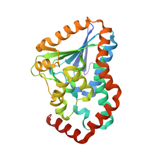
Domain swapping in the low-similarity isomerase/hydratase superfamily: the crystal structure of rat mitochondrial Delta3, Delta2-enoyl-CoA isomerase.

Hubbard, P.A.Yu, W.Schulz, H.Kim, J.J.

(2005) Protein Sci 14: 1545-1555

  • DOI: https://doi.org/10.1110/ps.041303705
  • Primary Citation of Related Structures:  
    1XX4

  • PubMed Abstract: 

    Two monofunctional Delta(3), Delta(2)-enoyl-CoA isomerases, one in mitochondria (mECI) and the other in both mitochondria and peroxisomes (pECI), belong to the low-similarity isomerase/hydratase superfamily. Both enzymes catalyze the movement of a double bond from C3 to C2 of an unsaturated acyl-CoA substrate for re-entry into the beta-oxidation pathway. Mutagenesis has shown that Glu165 of rat mECI is involved in catalysis; however, the putative catalytic residue in yeast pECI, Glu158, is not conserved in mECI. To elucidate whether Glu165 of mECI is correctly positioned for catalysis, the crystal structure of rat mECI has been solved. Crystal packing suggests the enzyme is trimeric, in contrast to other members of the superfamily, which appear crystallographically to be dimers of trimers. The polypeptide fold of mECI, like pECI, belongs to a subset of this superfamily in which the C-terminal domain of a given monomer interacts with its own N-terminal domain. This differs from that of crotonase and 1,4-dihydroxy-2-naphtoyl-CoA synthase, whose C-terminal domains are involved in domain swapping with an adjacent monomer. The structure confirms Glu165 as the putative catalytic acid/base, positioned to abstract the pro-R proton from C2 and reprotonate at C4 of the acyl chain. The large tunnel-shaped active site cavity observed in the mECI structure explains the relative substrate promiscuity in acyl-chain length and stereochemistry. Comparison with the crystal structure of pECI suggests the catalytic residues from both enzymes are spatially conserved but not in their primary structures, providing a powerful reminder of how catalytic residues cannot be determined solely by sequence alignments.


  • Organizational Affiliation

    Department of Biochemistry, Medical College of Wisconsin, Milwaukee, WI 53226, USA.


Macromolecules
Find similar proteins by:  (by identity cutoff)  |  3D Structure
Entity ID: 1
MoleculeChains Sequence LengthOrganismDetailsImage
3,2-trans-enoyl-CoA isomerase, mitochondrial261Rattus norvegicusMutation(s): 0 
Gene Names: Dci
EC: 5.3.3.8
UniProt
Find proteins for P23965 (Rattus norvegicus)
Explore P23965 
Go to UniProtKB:  P23965
Entity Groups  
Sequence Clusters30% Identity50% Identity70% Identity90% Identity95% Identity100% Identity
UniProt GroupP23965
Sequence Annotations
Expand
  • Reference Sequence
Small Molecules
Ligands 3 Unique
IDChains Name / Formula / InChI Key2D Diagram3D Interactions
BEN
Query on BEN

Download Ideal Coordinates CCD File 
H [auth A],
I [auth A],
J [auth A]
BENZAMIDINE
C7 H8 N2
PXXJHWLDUBFPOL-UHFFFAOYSA-N
PO4
Query on PO4

Download Ideal Coordinates CCD File 
B [auth A],
C [auth A],
D [auth A]
PHOSPHATE ION
O4 P
NBIIXXVUZAFLBC-UHFFFAOYSA-K
ZN
Query on ZN

Download Ideal Coordinates CCD File 
E [auth A],
F [auth A],
G [auth A]
ZINC ION
Zn
PTFCDOFLOPIGGS-UHFFFAOYSA-N
Experimental Data & Validation

Experimental Data

  • Method: X-RAY DIFFRACTION
  • Resolution: 2.20 Å
  • R-Value Free: 0.265 
  • R-Value Work: 0.239 
  • R-Value Observed: 0.245 
  • Space Group: I 41 3 2
Unit Cell:
Length ( Å )Angle ( ˚ )
a = 169.918α = 90
b = 169.918β = 90
c = 169.918γ = 90
Software Package:
Software NamePurpose
CNSrefinement
DENZOdata reduction
SCALEPACKdata scaling
MOLREPphasing

Structure Validation

View Full Validation Report


Entry History 

Deposition Data

Revision History  (Full details and data files)

  • Version 1.0: 2004-11-23
    Type: Initial release
  • Version 1.1: 2008-04-30
    Changes: Version format compliance
  • Version 1.2: 2011-07-13
    Changes: Derived calculations, Version format compliance
  • Version 1.3: 2011-11-16
    Changes: Atomic model
  • Version 2.0: 2021-08-04
    Changes: Atomic model, Data collection, Derived calculations, Non-polymer description, Structure summary
  • Version 2.1: 2024-02-14
    Changes: Data collection, Database references
  • Version 2.2: 2024-04-03
    Changes: Refinement description