1XUB

Structure and function of the phenazine biosynthetic protein PhzF from Pseudomonas fluorescens


Experimental Data Snapshot

  • Method: X-RAY DIFFRACTION
  • Resolution: 1.30 Å
  • R-Value Free: 0.160 
  • R-Value Work: 0.133 
  • R-Value Observed: 0.135 

wwPDB Validation   3D Report Full Report


This is version 1.4 of the entry. See complete history


Literature

Structure and function of the phenazine biosynthetic protein PhzF from Pseudomonas fluorescens

Blankenfeldt, W.Kuzin, A.P.Skarina, T.Korniyenko, Y.Tong, L.Bayer, P.Janning, P.Thomashow, L.S.Mavrodi, D.V.

(2004) Proc Natl Acad Sci U S A 101: 16431-16437

  • DOI: https://doi.org/10.1073/pnas.0407371101
  • Primary Citation of Related Structures:  
    1SDJ, 1U1V, 1U1W, 1U1X, 1XUA, 1XUB

  • PubMed Abstract: 

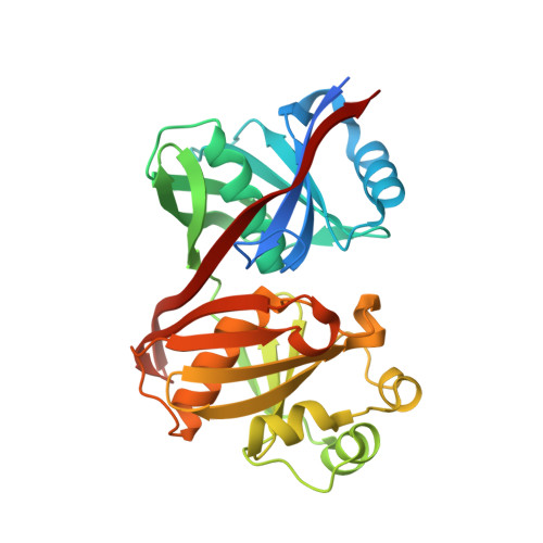
    Phenazines produced by Pseudomonas and Streptomyces spp. are heterocyclic nitrogen-containing metabolites with antibiotic, antitumor, and antiparasitic activity. The antibiotic properties of pyocyanin, produced by Pseudomonas aeruginosa, were recognized in the 1890s, although this blue phenazine is now known to be a virulence factor in human disease. Despite their biological significance, the biosynthesis of phenazines is not fully understood. Here we present structural and functional studies of PhzF, an enzyme essential for phenazine synthesis in Pseudomonas spp. PhzF shares topology with diaminopimelate epimerase DapF but lacks the same catalytic residues. The structure of PhzF in complex with its substrate, trans-2,3-dihydro-3-hydroxyanthranilic acid, suggests that it is an isomerase using the conserved glutamate E45 to abstract a proton from C3 of the substrate. The proton is returned to C1 of the substrate after rearrangement of the double-bond system, yielding an enol that converts to the corresponding ketone. PhzF is a dimer that may be bifunctional, providing a shielded cavity for ketone dimerization via double Schiff-base formation to produce the phenazine scaffold. Our proposed mechanism is supported by mass and NMR spectroscopy. The results are discussed in the context of related structures and protein sequences of unknown biochemical function.


  • Organizational Affiliation

    Max Planck Institute of Molecular Physiology, Otto-Hahn-Strasse 11, 44227 Dortmund, Germany.


Macromolecules
Find similar proteins by:  (by identity cutoff)  |  3D Structure
Entity ID: 1
MoleculeChains Sequence LengthOrganismDetailsImage
Phenazine biosynthesis protein phzF298Pseudomonas fluorescensMutation(s): 1 
UniProt
Find proteins for Q51792 (Pseudomonas fluorescens)
Explore Q51792 
Go to UniProtKB:  Q51792
Entity Groups  
Sequence Clusters30% Identity50% Identity70% Identity90% Identity95% Identity100% Identity
UniProt GroupQ51792
Sequence Annotations
Expand
  • Reference Sequence
Small Molecules
Ligands 1 Unique
IDChains Name / Formula / InChI Key2D Diagram3D Interactions
SO4
Query on SO4

Download Ideal Coordinates CCD File 
B [auth A]SULFATE ION
O4 S
QAOWNCQODCNURD-UHFFFAOYSA-L
Experimental Data & Validation

Experimental Data

  • Method: X-RAY DIFFRACTION
  • Resolution: 1.30 Å
  • R-Value Free: 0.160 
  • R-Value Work: 0.133 
  • R-Value Observed: 0.135 
  • Space Group: P 32 2 1
Unit Cell:
Length ( Å )Angle ( ˚ )
a = 56.128α = 90
b = 56.128β = 90
c = 154.423γ = 120
Software Package:
Software NamePurpose
REFMACrefinement
XDSdata reduction
XDSdata scaling

Structure Validation

View Full Validation Report


Entry History 

Deposition Data

Revision History  (Full details and data files)

  • Version 1.0: 2004-11-09
    Type: Initial release
  • Version 1.1: 2008-04-30
    Changes: Version format compliance
  • Version 1.2: 2011-07-13
    Changes: Version format compliance
  • Version 1.3: 2021-11-10
    Changes: Database references, Derived calculations
  • Version 1.4: 2023-10-25
    Changes: Data collection, Refinement description