1XSS

Semi-rational engineering of a green-emitting coral fluorescent protein into an efficient highlighter.


Experimental Data Snapshot

  • Method: X-RAY DIFFRACTION
  • Resolution: 1.60 Å
  • R-Value Free: 0.229 
  • R-Value Observed: 0.197 

wwPDB Validation   3D Report Full Report


This is version 2.0 of the entry. See complete history


Literature

Semi-rational engineering of a coral fluorescent protein into an efficient highlighter

Tsutsui, H.Karasawa, S.Shimizu, H.Nukina, N.Miyawaki, A.

(2005) EMBO Rep 6: 233-238

  • DOI: https://doi.org/10.1038/sj.embor.7400361
  • Primary Citation of Related Structures:  
    1XSS

  • PubMed Abstract: 

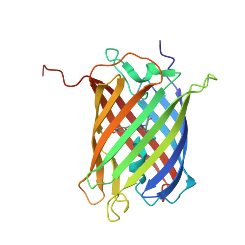
    Kaede is a natural photoconvertible fluorescent protein found in the coral Trachyphyllia geoffroyi. It contains a tripeptide, His 62-Tyr 63-Gly 64, which acts as a green chromophore that is photoconvertible to red following (ultra-) violet irradiation. Here, we report the molecular cloning and crystal structure determination of a new fluorescent protein, KikG, from the coral Favia favus, and its in vitro evolution conferring green-to-red photoconvertibility. Substitution of the His 62-Tyr 63-Gly 64 sequence into the native protein provided only negligible photoconversion. On the basis of the crystal structure, semi-rational mutagenesis of the amino acids surrounding the chromophore was performed, leading to the generation of an efficient highlighter, KikGR. Within mammalian cells, KikGR is more efficiently photoconverted and is several-fold brighter in both the green and red states than Kaede. In addition, KikGR was successfully photoconverted using two-photon excitation microscopy at 760 nm, ensuring optical cell labelling with better spatial discrimination in thick and highly scattering tissues.


  • Organizational Affiliation

    Laboratory for Cell Function Dynamics, Brain Science Institute, RIKEN, 2-1 Hirosawa, Wako-city, Saitama 351-0198, Japan.


Macromolecules
Find similar proteins by:  (by identity cutoff)  |  3D Structure
Entity ID: 1
MoleculeChains Sequence LengthOrganismDetailsImage
fluorescent protein
A, B
225Dipsastraea favusMutation(s): 1 
UniProt
Find proteins for Q53UG7 (Dipsastraea favus)
Explore Q53UG7 
Go to UniProtKB:  Q53UG7
Entity Groups  
Sequence Clusters30% Identity50% Identity70% Identity90% Identity95% Identity100% Identity
UniProt GroupQ53UG7
Sequence Annotations
Expand
  • Reference Sequence
Small Molecules
Modified Residues  1 Unique
IDChains TypeFormula2D DiagramParent
DYG
Query on DYG
A, B
L-PEPTIDE LINKINGC15 H15 N3 O6ASP, TYR, GLY
Experimental Data & Validation

Experimental Data

  • Method: X-RAY DIFFRACTION
  • Resolution: 1.60 Å
  • R-Value Free: 0.229 
  • R-Value Observed: 0.197 
  • Space Group: C 1 2 1
Unit Cell:
Length ( Å )Angle ( ˚ )
a = 96.74α = 90
b = 119.08β = 120.72
c = 49.29γ = 90
Software Package:
Software NamePurpose
MOSFLMdata reduction
SCALAdata scaling
MOLREPphasing
CNSrefinement
CCP4data scaling

Structure Validation

View Full Validation Report


Entry History 

Deposition Data

Revision History  (Full details and data files)

  • Version 1.0: 2005-03-15
    Type: Initial release
  • Version 1.1: 2008-04-30
    Changes: Version format compliance
  • Version 1.2: 2011-07-13
    Changes: Version format compliance
  • Version 1.3: 2011-11-16
    Changes: Atomic model
  • Version 2.0: 2023-11-15
    Changes: Atomic model, Data collection, Database references, Derived calculations