1XMZ

Crystal structure of the dark state of kindling fluorescent protein kfp from anemonia sulcata


Experimental Data Snapshot

  • Method: X-RAY DIFFRACTION
  • Resolution: 1.38 Å
  • R-Value Free: 0.191 
  • R-Value Work: 0.139 
  • R-Value Observed: 0.140 

wwPDB Validation   3D Report Full Report


This is version 2.0 of the entry. See complete history


Literature

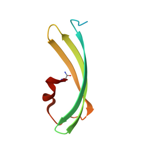
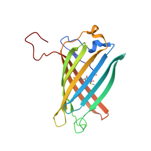
Kindling Fluorescent Protein from Anemonia sulcata: Dark-State Structure at 1.38 Resolution

Quillin, M.L.Anstrom, D.M.Shu, X.O'Leary, S.Kallio, K.Chudakov, D.M.Remington, S.J.

(2005) Biochemistry 44: 5774-5787

  • DOI: https://doi.org/10.1021/bi047644u
  • Primary Citation of Related Structures:  
    1XMZ

  • PubMed Abstract: 

    When the nonfluorescent chromoprotein asFP595 from Anemonia sulcata is subjected to sufficiently intense illumination near the absorbance maximum (lambda(abs)(max) = 568 nm), it undergoes a remarkable transition, termed "kindling", to a long-lived fluorescent state (lambda(em)(max) = 595 nm). In the dark recovery phase, the kindled state relaxes thermally on a time scale of seconds or can instantly be reverted upon illumination at 450 nm. The kindling phenomenon is enhanced by the Ala143 --> Gly point mutation, which slows the dark recovery time constant to 100 s at room temperature and increases the fluorescence quantum yield. To investigate the chemical nature of the chromophore and the possible role of chromophore isomerization in the kindling phenomenon, we determined the crystal structure of the "kindling fluorescent protein" asFP595-A143G (KFP) in the dark-adapted state at 1.38 A resolution and 100 K. The chromophore, derived from the Met63-Tyr64-Gly65 tripeptide, closely resembles that of the nonfluorescent chromoprotein Rtms5 in that the configuration is trans about the methylene bridge and there is substantial distortion from planarity. Unlike in Rtms5, in the native protein the polypeptide backbone is cleaved between Cys62 and Met63. The size and shape of the chromophore pocket suggest that the cis isomer of the chromophore could also be accommodated. Within the pocket, partially disordered His197 displays two conformations, which may constitute a binary switch that stabilizes different chromophore configurations. The energy barrier for thermal relaxation was found by Arrhenius plot analysis to be approximately 71 kJ/mol, somewhat higher than the value of approximately 55 kJ/mol observed for cis-trans isomerization of a model chromophore in solution.


  • Organizational Affiliation

    Department of Physics, Institute of Molecular Biology, University of Oregon, Eugene, Oregon 97403, USA.


Macromolecules
Find similar proteins by:  (by identity cutoff)  |  3D Structure
Entity ID: 1
MoleculeChains Sequence LengthOrganismDetailsImage
GFP-like non-fluorescent chromoprotein FP595 chain 1A,
C [auth B]
74Anemonia sulcataMutation(s): 0 
UniProt
Find proteins for Q9GZ28 (Anemonia sulcata)
Explore Q9GZ28 
Go to UniProtKB:  Q9GZ28
Entity Groups  
Sequence Clusters30% Identity50% Identity70% Identity90% Identity95% Identity100% Identity
UniProt GroupQ9GZ28
Sequence Annotations
Expand
  • Reference Sequence
Find similar proteins by:  (by identity cutoff)  |  3D Structure
Entity ID: 2
MoleculeChains Sequence LengthOrganismDetailsImage
GFP-like non-fluorescent chromoprotein FP595 chain 2B [auth C],
D
168Anemonia sulcataMutation(s): 2 
UniProt
Find proteins for Q9GZ28 (Anemonia sulcata)
Explore Q9GZ28 
Go to UniProtKB:  Q9GZ28
Entity Groups  
Sequence Clusters30% Identity50% Identity70% Identity90% Identity95% Identity100% Identity
UniProt GroupQ9GZ28
Sequence Annotations
Expand
  • Reference Sequence
Experimental Data & Validation

Experimental Data

  • Method: X-RAY DIFFRACTION
  • Resolution: 1.38 Å
  • R-Value Free: 0.191 
  • R-Value Work: 0.139 
  • R-Value Observed: 0.140 
  • Space Group: C 2 2 21
Unit Cell:
Length ( Å )Angle ( ˚ )
a = 76.52α = 90
b = 125.4β = 90
c = 92.75γ = 90
Software Package:
Software NamePurpose
HKL-2000data collection
SCALEPACKdata scaling
EPMRphasing
SHELXL-97refinement
HKL-2000data reduction

Structure Validation

View Full Validation Report


Entry History 

Deposition Data

Revision History  (Full details and data files)

  • Version 1.0: 2005-04-19
    Type: Initial release
  • Version 1.1: 2008-04-30
    Changes: Version format compliance
  • Version 1.2: 2011-07-13
    Changes: Derived calculations, Version format compliance
  • Version 1.3: 2021-10-20
    Changes: Database references, Derived calculations
  • Version 1.4: 2023-08-23
    Changes: Data collection, Refinement description
  • Version 2.0: 2024-02-28
    Changes: Advisory, Atomic model, Data collection, Database references, Derived calculations, Polymer sequence, Source and taxonomy, Structure summary