1XMK

The Crystal structure of the Zb domain from the RNA editing enzyme ADAR1


Experimental Data Snapshot

  • Method: X-RAY DIFFRACTION
  • Resolution: 0.97 Å
  • R-Value Free: 0.183 
  • R-Value Observed: 0.145 

wwPDB Validation   3D Report Full Report


This is version 1.4 of the entry. See complete history


Literature

The Crystal Structure of the Z[beta] Domain of the RNA-editing Enzyme ADAR1 Reveals Distinct Conserved Surfaces Among Z-domains.

Athanasiadis, A.Placido, D.Maas, S.Brown II, B.A.Lowenhaupt, K.Rich, A.

(2005) J Mol Biol 351: 496-507

  • DOI: https://doi.org/10.1016/j.jmb.2005.06.028
  • Primary Citation of Related Structures:  
    1XMK

  • PubMed Abstract: 

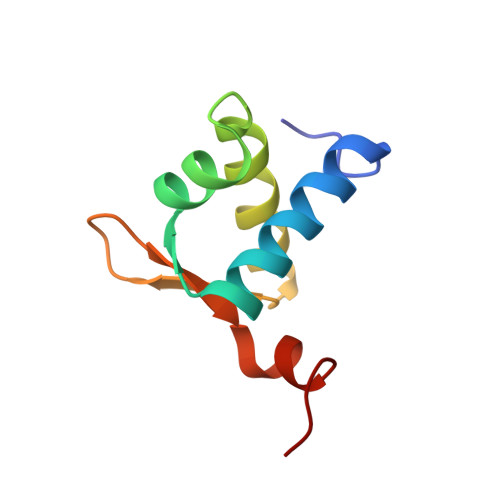
    The Zalpha domains represent a growing subfamily of the winged helix-turn-helix (HTH) domain family whose members share a remarkable ability to bind specifically to Z-DNA and/or Z-RNA. They have been found exclusively in proteins involved in interferon response and, while their importance in determining pox viral pathogenicity has been demonstrated, their actual target and biological role remain obscure. Cellular proteins containing Zalpha domains bear a second homologous domain termed Zbeta, which appears to lack the ability to bind left-handed nucleic acids. Here, we present the crystal structure of the Zbeta domain from the human double-stranded RNA adenosine deaminase ADAR1 at 0.97 A, determined by single isomorphous replacement including anomalous scattering. Zbeta maintains a winged-HTH fold with the addition of a C-terminal helix. Mapping of the Zbeta conservation profile on the Zbeta surface reveals a new conserved surface formed partly by the terminal helix 4, involved in metal binding and dimerization and absent from Zalpha domains. Our results show how two domains similar in fold may have evolved into different functional entities even in the context of the same protein.


  • Organizational Affiliation

    Department of Biology, Massachusetts Institute of Technology, 77 Massachusetts Avenue, Cambridge, MA 02139, USA. alekosathanasiadis@hotmail.com


Macromolecules
Find similar proteins by:  (by identity cutoff)  |  3D Structure
Entity ID: 1
MoleculeChains Sequence LengthOrganismDetailsImage
Double-stranded RNA-specific adenosine deaminase79Homo sapiensMutation(s): 0 
Gene Names: ADARADAR1DSRADIFI4
EC: 3.5.4
UniProt & NIH Common Fund Data Resources
Find proteins for P55265 (Homo sapiens)
Explore P55265 
Go to UniProtKB:  P55265
PHAROS:  P55265
GTEx:  ENSG00000160710 
Entity Groups  
Sequence Clusters30% Identity50% Identity70% Identity90% Identity95% Identity100% Identity
UniProt GroupP55265
Sequence Annotations
Expand
  • Reference Sequence
Experimental Data & Validation

Experimental Data

  • Method: X-RAY DIFFRACTION
  • Resolution: 0.97 Å
  • R-Value Free: 0.183 
  • R-Value Observed: 0.145 
  • Space Group: P 21 21 21
Unit Cell:
Length ( Å )Angle ( ˚ )
a = 35.557α = 90
b = 43.526β = 90
c = 45.471γ = 90
Software Package:
Software NamePurpose
SHELXrefinement
MOSFLMdata reduction
CCP4data scaling
SHARPphasing
SHELXLrefinement

Structure Validation

View Full Validation Report


Entry History 

Deposition Data

Revision History  (Full details and data files)

  • Version 1.0: 2005-08-02
    Type: Initial release
  • Version 1.1: 2008-04-30
    Changes: Version format compliance
  • Version 1.2: 2011-07-13
    Changes: Version format compliance
  • Version 1.3: 2018-01-24
    Changes: Database references
  • Version 1.4: 2024-02-14
    Changes: Data collection, Database references, Derived calculations