1XGR

Structure for antibody HyHEL-63 Y33I mutant complexed with hen egg lysozyme


Experimental Data Snapshot

  • Method: X-RAY DIFFRACTION
  • Resolution: 2.10 Å
  • R-Value Free: 0.304 
  • R-Value Work: 0.250 
  • R-Value Observed: 0.250 

wwPDB Validation   3D Report Full Report


This is version 1.3 of the entry. See complete history


Literature

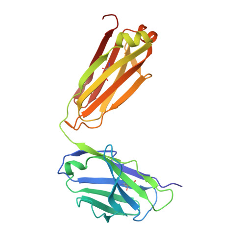
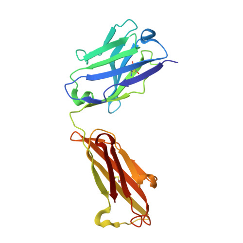
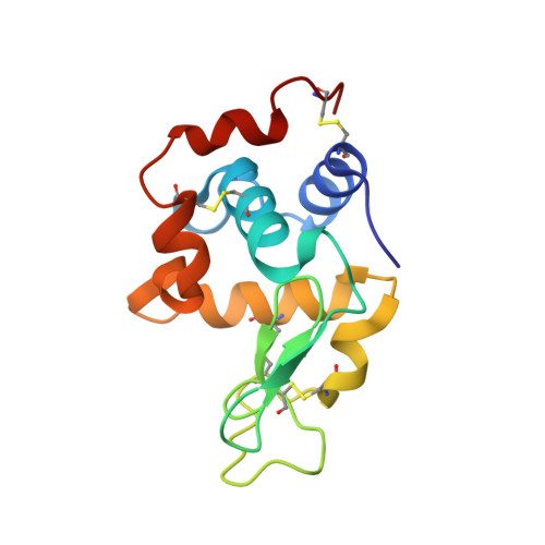
Magnitude of the hydrophobic effect at central versus peripheral sites in protein-protein interfaces

Li, Y.Huang, Y.Swaminathan, C.P.Smith-Gill, S.J.Mariuzza, R.A.

(2005) Structure 13: 297-307

  • DOI: https://doi.org/10.1016/j.str.2004.12.012
  • Primary Citation of Related Structures:  
    1XGP, 1XGQ, 1XGR, 1XGT, 1XGU

  • PubMed Abstract: 

    Hydrophobic interactions are essential for stabilizing protein-protein complexes, whose interfaces generally consist of a central cluster of hot spot residues surrounded by less important peripheral residues. According to the O-ring hypothesis, a condition for high affinity binding is solvent exclusion from interacting residues. This hypothesis predicts that the hydrophobicity at the center is significantly greater than at the periphery, which we estimated at 21 cal mol(-1) A(-2). To measure the hydrophobicity at the center, structures of an antigen-antibody complex where a buried phenylalanine was replaced by smaller hydrophobic residues were determined. By correlating structural changes with binding free energies, we estimate the hydrophobicity at this central site to be 46 cal mol(-1) A(-2), twice that at the periphery. This context dependence of the hydrophobic effect explains the clustering of hot spots at interface centers and has implications for hot spot prediction and the design of small molecule inhibitors.


  • Organizational Affiliation

    Center for Advanced Research in Biotechnology, W.M. Keck Laboratory for Structural Biology, University of Maryland Biotechnology Institute, 9600 Gudelsky Drive, Rockville, Maryland 20859, USA.


Macromolecules
Find similar proteins by:  (by identity cutoff)  |  3D Structure
Entity ID: 1
MoleculeChains Sequence LengthOrganismDetailsImage
antibody kappa light chain215Mus musculusMutation(s): 0 
Entity Groups  
Sequence Clusters30% Identity50% Identity70% Identity90% Identity95% Identity100% Identity
Sequence Annotations
Expand
  • Reference Sequence
Find similar proteins by:  (by identity cutoff)  |  3D Structure
Entity ID: 2
MoleculeChains Sequence LengthOrganismDetailsImage
antibody kappa heavy chain210Mus musculusMutation(s): 0 
Entity Groups  
Sequence Clusters30% Identity50% Identity70% Identity90% Identity95% Identity100% Identity
Sequence Annotations
Expand
  • Reference Sequence
Find similar proteins by:  (by identity cutoff)  |  3D Structure
Entity ID: 3
MoleculeChains Sequence LengthOrganismDetailsImage
Lysozyme C129Gallus gallusMutation(s): 0 
EC: 3.2.1.17
UniProt
Find proteins for P00698 (Gallus gallus)
Explore P00698 
Go to UniProtKB:  P00698
Entity Groups  
Sequence Clusters30% Identity50% Identity70% Identity90% Identity95% Identity100% Identity
UniProt GroupP00698
Sequence Annotations
Expand
  • Reference Sequence
Experimental Data & Validation

Experimental Data

  • Method: X-RAY DIFFRACTION
  • Resolution: 2.10 Å
  • R-Value Free: 0.304 
  • R-Value Work: 0.250 
  • R-Value Observed: 0.250 
  • Space Group: P 42 21 2
Unit Cell:
Length ( Å )Angle ( ˚ )
a = 90.316α = 90
b = 90.316β = 90
c = 150.592γ = 90
Software Package:
Software NamePurpose
CNSrefinement

Structure Validation

View Full Validation Report


Entry History 

Deposition Data

Revision History  (Full details and data files)

  • Version 1.0: 2005-09-06
    Type: Initial release
  • Version 1.1: 2007-10-21
    Changes: Version format compliance
  • Version 1.2: 2011-07-13
    Changes: Version format compliance
  • Version 1.3: 2013-10-02
    Changes: Source and taxonomy, Structure summary