1XDJ

Crystal Structure of T. maritima Cobalamin-Independent Methionine Synthase complexed with Zn2+ and Homocysteine


Experimental Data Snapshot

  • Method: X-RAY DIFFRACTION
  • Resolution: 2.20 Å
  • R-Value Free: 0.268 
  • R-Value Work: 0.227 
  • R-Value Observed: 0.227 

wwPDB Validation   3D Report Full Report


This is version 1.4 of the entry. See complete history


Literature

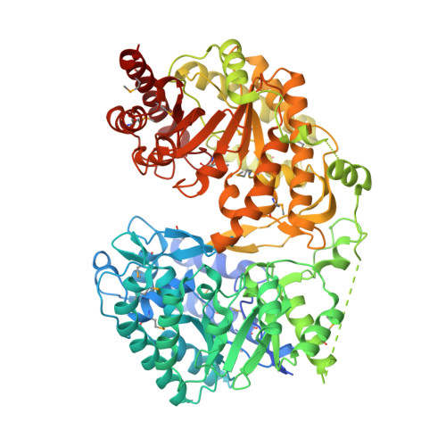
Cobalamin-independent methionine synthase (MetE): a face-to-face double barrel that evolved by gene duplication

Pejchal, R.Ludwig, M.L.

(2005) PLoS Biol 3: e31

  • DOI: https://doi.org/10.1371/journal.pbio.0030031
  • Primary Citation of Related Structures:  
    1T7L, 1XDJ, 1XPG, 1XR2

  • PubMed Abstract: 

    Cobalamin-independent methionine synthase (MetE) catalyzes the transfer of a methyl group from methyltetrahydrofolate to L-homocysteine (Hcy) without using an intermediate methyl carrier. Although MetE displays no detectable sequence homology with cobalamin-dependent methionine synthase (MetH), both enzymes require zinc for activation and binding of Hcy. Crystallographic analyses of MetE from T. maritima reveal an unusual dual-barrel structure in which the active site lies between the tops of the two (betaalpha)(8) barrels. The fold of the N-terminal barrel confirms that it has evolved from the C-terminal polypeptide by gene duplication; comparisons of the barrels provide an intriguing example of homologous domain evolution in which binding sites are obliterated. The C-terminal barrel incorporates the zinc ion that binds and activates Hcy. The zinc-binding site in MetE is distinguished from the (Cys)(3)Zn site in the related enzymes, MetH and betaine-homocysteine methyltransferase, by its position in the barrel and by the metal ligands, which are histidine, cysteine, glutamate, and cysteine in the resting form of MetE. Hcy associates at the face of the metal opposite glutamate, which moves away from the zinc in the binary E.Hcy complex. The folate substrate is not intimately associated with the N-terminal barrel; instead, elements from both barrels contribute binding determinants in a binary complex in which the folate substrate is incorrectly oriented for methyl transfer. Atypical locations of the Hcy and folate sites in the C-terminal barrel presumably permit direct interaction of the substrates in a ternary complex. Structures of the binary substrate complexes imply that rearrangement of folate, perhaps accompanied by domain rearrangement, must occur before formation of a ternary complex that is competent for methyl transfer.


  • Organizational Affiliation

    Department of Biological Chemistry, University of Michigan, Ann Arbor, USA.


Macromolecules
Find similar proteins by:  (by identity cutoff)  |  3D Structure
Entity ID: 1
MoleculeChains Sequence LengthOrganismDetailsImage
5-methyltetrahydropteroyltriglutamate--homocysteine methyltransferase
A, B
766Thermotoga maritimaMutation(s): 17 
Gene Names: metE
EC: 2.1.1.14
UniProt
Find proteins for Q9X112 (Thermotoga maritima (strain ATCC 43589 / DSM 3109 / JCM 10099 / NBRC 100826 / MSB8))
Explore Q9X112 
Go to UniProtKB:  Q9X112
Entity Groups  
Sequence Clusters30% Identity50% Identity70% Identity90% Identity95% Identity100% Identity
UniProt GroupQ9X112
Sequence Annotations
Expand
  • Reference Sequence
Small Molecules
Ligands 4 Unique
IDChains Name / Formula / InChI Key2D Diagram3D Interactions
HCS
Query on HCS

Download Ideal Coordinates CCD File 
E [auth A],
F [auth A]
2-AMINO-4-MERCAPTO-BUTYRIC ACID
C4 H9 N O2 S
FFFHZYDWPBMWHY-VKHMYHEASA-N
MRY
Query on MRY

Download Ideal Coordinates CCD File 
G [auth A],
J [auth B]
MESO-ERYTHRITOL
C4 H10 O4
UNXHWFMMPAWVPI-ZXZARUISSA-N
SO4
Query on SO4

Download Ideal Coordinates CCD File 
C [auth A],
H [auth B]
SULFATE ION
O4 S
QAOWNCQODCNURD-UHFFFAOYSA-L
ZN
Query on ZN

Download Ideal Coordinates CCD File 
D [auth A],
I [auth B]
ZINC ION
Zn
PTFCDOFLOPIGGS-UHFFFAOYSA-N
Modified Residues  1 Unique
IDChains TypeFormula2D DiagramParent
MSE
Query on MSE
A, B
L-PEPTIDE LINKINGC5 H11 N O2 SeMET
Experimental Data & Validation

Experimental Data

  • Method: X-RAY DIFFRACTION
  • Resolution: 2.20 Å
  • R-Value Free: 0.268 
  • R-Value Work: 0.227 
  • R-Value Observed: 0.227 
  • Space Group: P 21 21 2
Unit Cell:
Length ( Å )Angle ( ˚ )
a = 167.053α = 90
b = 159.366β = 90
c = 65.639γ = 90
Software Package:
Software NamePurpose
CNSrefinement
MAR345data collection
SCALEPACKdata scaling
EPMRphasing

Structure Validation

View Full Validation Report


Entry History 

Deposition Data

Revision History  (Full details and data files)

  • Version 1.0: 2005-03-01
    Type: Initial release
  • Version 1.1: 2007-10-21
    Changes: Version format compliance
  • Version 1.2: 2011-07-13
    Changes: Version format compliance
  • Version 1.3: 2017-10-11
    Changes: Refinement description
  • Version 1.4: 2024-04-03
    Changes: Data collection, Database references, Derived calculations, Structure summary