1XAE

Crystal structure of wild type yellow fluorescent protein zFP538 from Zoanthus


Experimental Data Snapshot

  • Method: X-RAY DIFFRACTION
  • Resolution: 2.70 Å
  • R-Value Free: 0.255 
  • R-Value Work: 0.205 
  • R-Value Observed: 0.206 

wwPDB Validation   3D Report Full Report


This is version 1.5 of the entry. See complete history


Literature

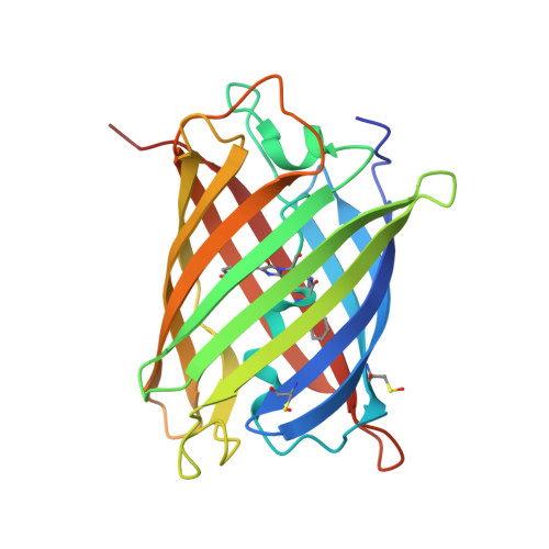
zFP538, a yellow-fluorescent protein from Zoanthus, contains a novel three-ring chromophore.

Remington, S.J.Wachter, R.M.Yarbrough, D.K.Branchaud, B.Anderson, D.C.Kallio, K.Lukyanov, K.A.

(2005) Biochemistry 44: 202-212

  • DOI: https://doi.org/10.1021/bi048383r
  • Primary Citation of Related Structures:  
    1XA9, 1XAE

  • PubMed Abstract: 

    Crystal structures of the tetrameric yellow-fluorescent protein zFP538 from the button polyp Zoanthus sp. and a green-emitting mutant (K66M) are presented. The atomic models have been refined at 2.7 and 2.5 A resolution, with final crystallographic R factors of 0.206 (R(free) = 0.255) and 0.190 (R(free) = 0.295), respectively, and have excellent stereochemistry. The fold of the protomer is very similar to that of green (GFP) and red (DsRed) fluorescent proteins; however, evidence from crystallography and mass spectrometry suggests that zFP538 contains a three-ring chromophore derived from that of GFP. The yellow-emitting species (lambda(em)(max) = 538 nm) is proposed to result from a transimination reaction in which a transiently appearing DsRed-like acylimine is attacked by the terminal amino group of lysine 66 to form a new six-membered ring, cleaving the polypeptide backbone at the 65-66 position. This extends the chromophore conjugation by an additional double bond compared to GFP, lowering the absorption and emission frequencies. Substitution of lysine 66 with aspartate or glutamate partially converts zFP538 into a red-fluorescent protein, providing additional support for an acylimine intermediate. The diverse and unexpected roles of the side chain at position 66 give new insight into the chemistry of chromophore maturation in the extended family of GFP-like proteins.


  • Organizational Affiliation

    Institute of Molecular Biology, Department of Physics, University of Oregon, Eugene, Oregon 97403-1229, USA. jremington@uoxray.uoregon.edu


Macromolecules
Find similar proteins by:  (by identity cutoff)  |  3D Structure
Entity ID: 1
MoleculeChains Sequence LengthOrganismDetailsImage
fluorescent protein FP538
A, B
229Zoanthus sp.Mutation(s): 1 
UniProt
Find proteins for Q9U6Y4 (Zoanthus sp.)
Explore Q9U6Y4 
Go to UniProtKB:  Q9U6Y4
Entity Groups  
Sequence Clusters30% Identity50% Identity70% Identity90% Identity95% Identity100% Identity
UniProt GroupQ9U6Y4
Sequence Annotations
Expand
  • Reference Sequence
Small Molecules
Modified Residues  3 Unique
IDChains TypeFormula2D DiagramParent
CH7
Query on CH7
A, B
L-PEPTIDE LINKINGC17 H17 N3 O4LYS, TYR, GLY
CSO
Query on CSO
A, B
L-PEPTIDE LINKINGC3 H7 N O3 SCYS
NFA
Query on NFA
A, B
L-PEPTIDE LINKINGC9 H12 N2 OPHE
Experimental Data & Validation

Experimental Data

  • Method: X-RAY DIFFRACTION
  • Resolution: 2.70 Å
  • R-Value Free: 0.255 
  • R-Value Work: 0.205 
  • R-Value Observed: 0.206 
  • Space Group: P 31 2 1
Unit Cell:
Length ( Å )Angle ( ˚ )
a = 121.16α = 90
b = 121.16β = 90
c = 111.69γ = 120
Software Package:
Software NamePurpose
DENZOdata reduction
SCALEPACKdata scaling
EPMRphasing
TNTrefinement

Structure Validation

View Full Validation Report


Entry History 

Deposition Data

Revision History  (Full details and data files)

  • Version 1.0: 2005-02-08
    Type: Initial release
  • Version 1.1: 2008-04-30
    Changes: Version format compliance
  • Version 1.2: 2011-07-13
    Changes: Version format compliance
  • Version 1.3: 2018-04-04
    Changes: Data collection
  • Version 1.4: 2023-08-23
    Changes: Data collection, Database references, Derived calculations, Refinement description
  • Version 1.5: 2023-11-15
    Changes: Data collection