1XA2

Cobalt hexammine induced tautomeric shift in Z-DNA: the structure of d(CGCGCA).d(TGCGCG) in two crystal forms


Experimental Data Snapshot

  • Method: X-RAY DIFFRACTION
  • Resolution: 1.71 Å
  • R-Value Free: 0.240 
  • R-Value Work: 0.208 
  • R-Value Observed: 0.210 

wwPDB Validation   3D Report Full Report


This is version 1.5 of the entry. See complete history


Literature

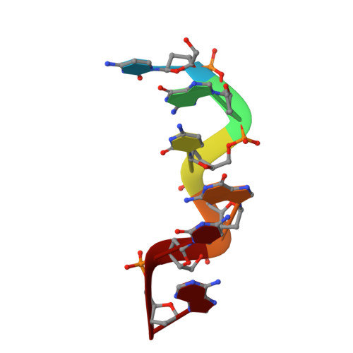
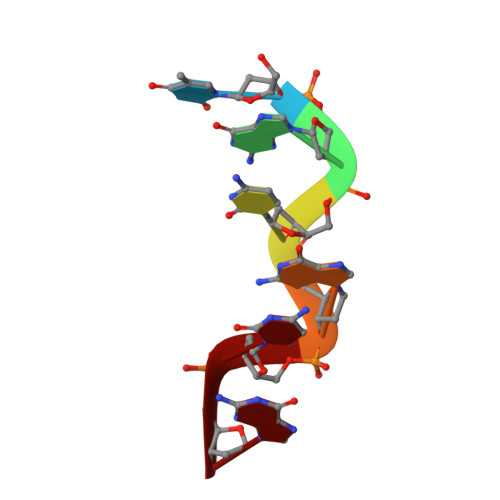
Cobalt hexammine induced tautomeric shift in Z-DNA: the structure of d(CGCGCA)*d(TGCGCG) in two crystal forms.

Thiyagarajan, S.Rajan, S.S.Gautham, N.

(2004) Nucleic Acids Res 32: 5945-5953

  • DOI: https://doi.org/10.1093/nar/gkh919
  • Primary Citation of Related Structures:  
    1XA2, 1XAM

  • PubMed Abstract: 

    We report here the crystal structure of the DNA hexamer duplex d(CGCGCA)*d(TGCGCG) at 1.71 A resolution. The crystals, in orthorhombic space group, were grown in the presence of cobalt hexammine, a known inducer of the left-handed Z form of DNA. The interaction of this ion with the DNA helix results in a change of the adenine base from the common amino tautomeric form to the imino tautomer. Consequently the A:T base pair is disrupted from the normal Watson-Crick base pairing to a 'wobble' like base pairing. This change is accommodated easily within the helix, and the helical parameters are those expected for Z-DNA. When the cobalt hexammine concentration is decreased slightly in the crystallization conditions, the duplex crystallizes in a different, hexagonal space group, with two hexamer duplexes in the asymmetric unit. One of these is situated on a crystallographic 6-fold screw axis, leading to disorder. The tautomeric shift is not observed in this space group. We show that the change in inter-helix interactions that lead to the two different space groups probably arise from the small decrease in ion concentration, and consequently disordered positions for the ion.


  • Organizational Affiliation

    Department of Crystallography and Biophysics, University of Madras, Guindy Campus, Chennai 600 025, India.


Macromolecules

Find similar nucleic acids by:  Sequence   |   3D Structure  

Entity ID: 1
MoleculeChains LengthOrganismImage
5'-D(*CP*GP*CP*GP*CP*A)-3'6N/A
Sequence Annotations
Expand
  • Reference Sequence

Find similar nucleic acids by:  Sequence   |   3D Structure  

Entity ID: 2
MoleculeChains LengthOrganismImage
5'-D(*TP*GP*CP*GP*CP*G)-3'6N/A
Sequence Annotations
Expand
  • Reference Sequence
Small Molecules
Ligands 1 Unique
IDChains Name / Formula / InChI Key2D Diagram3D Interactions
NCO
Query on NCO

Download Ideal Coordinates CCD File 
C [auth A]COBALT HEXAMMINE(III)
Co H18 N6
DYLMFCCYOUSRTK-UHFFFAOYSA-N
Experimental Data & Validation

Experimental Data

  • Method: X-RAY DIFFRACTION
  • Resolution: 1.71 Å
  • R-Value Free: 0.240 
  • R-Value Work: 0.208 
  • R-Value Observed: 0.210 
  • Space Group: P 21 21 21
Unit Cell:
Length ( Å )Angle ( ˚ )
a = 17.976α = 90
b = 30.926β = 90
c = 44.625γ = 90
Software Package:
Software NamePurpose
REFMACrefinement
MAR345data collection
AUTOMARdata reduction
AMoREphasing

Structure Validation

View Full Validation Report


Entry History 

Deposition Data

Revision History  (Full details and data files)

  • Version 1.0: 2004-11-16
    Type: Initial release
  • Version 1.1: 2008-04-30
    Changes: Version format compliance
  • Version 1.2: 2011-07-13
    Changes: Version format compliance
  • Version 1.3: 2017-10-11
    Changes: Refinement description
  • Version 1.4: 2024-02-14
    Changes: Data collection, Database references, Derived calculations
  • Version 1.5: 2024-04-03
    Changes: Refinement description