1X8S

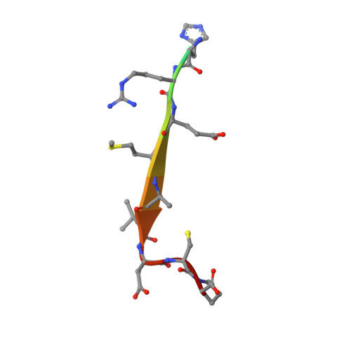
Structure of the Par-6 PDZ domain with a Pals1 internal ligand


Experimental Data Snapshot

  • Method: X-RAY DIFFRACTION
  • Resolution: 2.50 Å
  • R-Value Free: 0.254 
  • R-Value Work: 0.216 
  • R-Value Observed: 0.217 

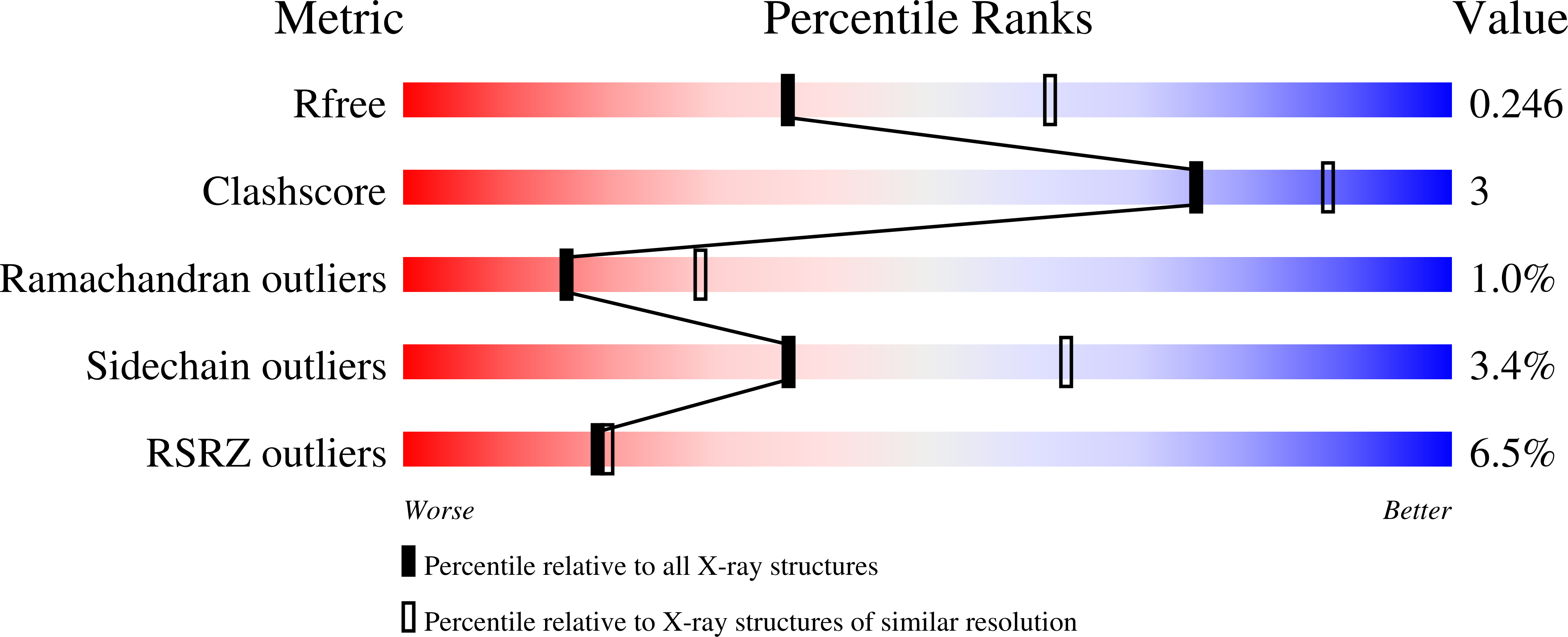
wwPDB Validation   3D Report Full Report


This is version 1.3 of the entry. See complete history


Literature

Internal recognition through PDZ domain plasticity in the Par-6-Pals1 complex.

Penkert, R.R.Divittorio, H.M.Prehoda, K.E.

(2004) Nat Struct Mol Biol 11: 1122-1127

  • DOI: https://doi.org/10.1038/nsmb839
  • Primary Citation of Related Structures:  
    1X8S

  • PubMed Abstract: 

    PDZ protein interaction domains are typically selective for C-terminal ligands, but non-C-terminal, 'internal' ligands have also been identified. The PDZ domain from the cell polarity protein Par-6 binds C-terminal ligands and an internal sequence from the protein Pals1/Stardust. The structure of the Pals1-Par-6 PDZ complex reveals that the PDZ ligand-binding site is deformed to allow for internal binding. Whereas binding of the Rho GTPase Cdc42 to a CRIB domain adjacent to the Par-6 PDZ regulates binding of C-terminal ligands, the conformational change that occurs upon binding of Pals1 renders its binding independent of Cdc42. These results suggest a mechanism by which the requirement for a C terminus can be readily bypassed by PDZ ligands and reveal a complex set of cooperative and competitive interactions in Par-6 that are likely to be important for cell polarity regulation.


  • Organizational Affiliation

    Institute of Molecular Biology and Department of Chemistry, University of Oregon, Eugene, Oregon 97403, USA.


Macromolecules
Find similar proteins by:  (by identity cutoff)  |  3D Structure
Entity ID: 1
MoleculeChains Sequence LengthOrganismDetailsImage
CG5884-PA102Drosophila melanogasterMutation(s): 0 
Gene Names: PAR-6
UniProt
Find proteins for O97111 (Drosophila melanogaster)
Explore O97111 
Go to UniProtKB:  O97111
Entity Groups  
Sequence Clusters30% Identity50% Identity70% Identity90% Identity95% Identity100% Identity
UniProt GroupO97111
Sequence Annotations
Expand
  • Reference Sequence

Find similar proteins by:  Sequence   |   3D Structure  

Entity ID: 2
MoleculeChains Sequence LengthOrganismDetailsImage
Pals1 peptide12N/AMutation(s): 0 
Entity Groups  
Sequence Clusters30% Identity50% Identity70% Identity90% Identity95% Identity100% Identity
Sequence Annotations
Expand
  • Reference Sequence
Experimental Data & Validation

Experimental Data

  • Method: X-RAY DIFFRACTION
  • Resolution: 2.50 Å
  • R-Value Free: 0.254 
  • R-Value Work: 0.216 
  • R-Value Observed: 0.217 
  • Space Group: H 3
Unit Cell:
Length ( Å )Angle ( ˚ )
a = 63.122α = 90
b = 63.122β = 90
c = 99.276γ = 120
Software Package:
Software NamePurpose
REFMACrefinement
SCALEPACKdata scaling
AMoREphasing

Structure Validation

View Full Validation Report


Entry History 

Deposition Data

Revision History  (Full details and data files)

  • Version 1.0: 2004-12-07
    Type: Initial release
  • Version 1.1: 2008-04-30
    Changes: Version format compliance
  • Version 1.2: 2011-07-13
    Changes: Advisory, Version format compliance
  • Version 1.3: 2024-02-14
    Changes: Data collection, Database references