


Crystal structure of ubch5c
Nakanishi, M., Teshima, N., Mizushima, T., Murata, S., Tanaka, K., Yamane, T.To be published.
Experimental Data Snapshot
wwPDB Validation 3D Report Full Report

Entity ID: 1 | |||||
|---|---|---|---|---|---|
| Molecule | Chains | Sequence Length | Organism | Details | Image |
| Ubiquitin-conjugating enzyme E2 D3 | 155 | Homo sapiens | Mutation(s): 0 EC: 6.3.2.19 | 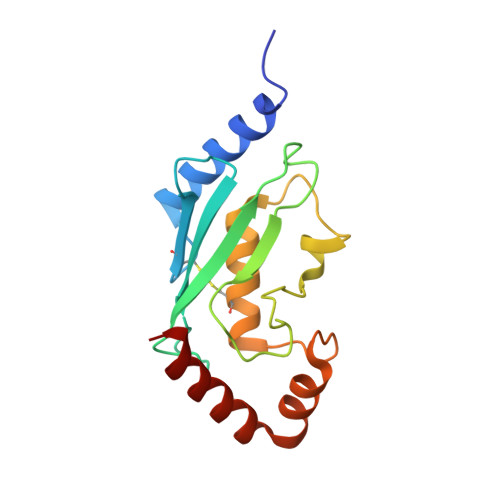 | |
UniProt & NIH Common Fund Data Resources | |||||
Find proteins for P61077 (Homo sapiens) Explore P61077 Go to UniProtKB: P61077 | |||||
PHAROS: P61077 GTEx: ENSG00000109332 | |||||
Entity Groups | |||||
| Sequence Clusters | 30% Identity50% Identity70% Identity90% Identity95% Identity100% Identity | ||||
| UniProt Group | P61077 | ||||
Sequence AnnotationsExpand | |||||
| |||||
| Length ( Å ) | Angle ( ˚ ) |
|---|---|
| a = 61.998 | α = 90 |
| b = 44.334 | β = 93.36 |
| c = 97.584 | γ = 90 |
| Software Name | Purpose |
|---|---|
| REFMAC | refinement |
| DENZO | data reduction |
| SCALEPACK | data scaling |
| CNS | phasing |
RCSB PDB (citation) is hosted by
RCSB PDB is a member of the

