1W8C

CO-CRYSTAL STRUCTURE OF 6-CYCLOHEXYLMETHOXY-8-ISOPROPYL-9H-PURIN-2- YLAMINE AND MONOMERIC CDK2


Experimental Data Snapshot

  • Method: X-RAY DIFFRACTION
  • Resolution: 2.05 Å
  • R-Value Free: 0.256 
  • R-Value Work: 0.196 
  • R-Value Observed: 0.199 

wwPDB Validation   3D Report Full Report


Ligand Structure Quality Assessment 


This is version 1.5 of the entry. See complete history


Literature

8-Substituted O6-Cyclohexylmethylguanine Cdk2 Inhibitors; Using Structure-Based Inhibitor Design to Optimise an Alternative Binding Mode.

Carbain, B.Paterson, D.J.Anscombe, E.Campbell-Dexter, A.Cano, C.Echalier, A.Endicott, J.Golding, B.T.Haggerty, K.Hardcastle, I.R.Jewsbury, P.J.Newell, D.R.Noble, M.Roche, C.Wang, L.Griffin, R.J.

(2014) J Med Chem 57: 56

  • DOI: https://doi.org/10.1021/jm401555v
  • Primary Citation of Related Structures:  
    1W8C, 4CFM, 4CFN, 4CFU, 4CFV, 4CFW, 4CFX

  • PubMed Abstract: 

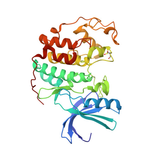
    Evaluation of the effects of purine C-8 substitution within a series of CDK1/2-selective O(6)-cyclohexylmethylguanine derivatives revealed that potency decreases initially with increasing size of the alkyl substituent. Structural analysis showed that C-8 substitution is poorly tolerated, and to avoid unacceptable steric interactions, these compounds adopt novel binding modes. Thus, 2-amino-6-cyclohexylmethoxy-8-isopropyl-9H-purine adopts a "reverse" binding mode where the purine backbone has flipped 180°. This provided a novel lead chemotype from which we have designed more potent CDK2 inhibitors using, in the first instance, quantum mechanical energy calculations. Introduction of an ortho-tolyl or ortho-chlorophenyl group at the purine C-8 position restored the potency of these "reverse" binding mode inhibitors to that of the parent 2-amino-6-cyclohexylmethoxy-9H-purine. By contrast, the corresponding 8-(2-methyl-3-sulfamoylphenyl)-purine derivative exhibited submicromolar CDK2-inhibitory activity by virtue of engineered additional interactions with Asp86 and Lys89 in the reversed binding mode, as confirmed by X-ray crystallography.


  • Organizational Affiliation

    Department of Biochemistry, University of Oxford , South Parks Road, Oxford OX1 3QU, U.K.


Macromolecules
Find similar proteins by:  (by identity cutoff)  |  3D Structure
Entity ID: 1
MoleculeChains Sequence LengthOrganismDetailsImage
CELL DIVISION PROTEIN KINASE 2299Homo sapiensMutation(s): 0 
EC: 2.7.1.37
UniProt & NIH Common Fund Data Resources
Find proteins for P24941 (Homo sapiens)
Explore P24941 
Go to UniProtKB:  P24941
PHAROS:  P24941
GTEx:  ENSG00000123374 
Entity Groups  
Sequence Clusters30% Identity50% Identity70% Identity90% Identity95% Identity100% Identity
UniProt GroupP24941
Sequence Annotations
Expand
  • Reference Sequence
Small Molecules
Ligands 1 Unique
IDChains Name / Formula / InChI Key2D Diagram3D Interactions
N69
Query on N69

Download Ideal Coordinates CCD File 
B [auth A]6-(CYCLOHEXYLMETHOXY)-8-ISOPROPYL-9H-PURIN-2-AMINE
C15 H23 N5 O
JABXNQUXGJTKRO-UHFFFAOYSA-N
Modified Residues  1 Unique
IDChains TypeFormula2D DiagramParent
CSD
Query on CSD
A
L-PEPTIDE LINKINGC3 H7 N O4 SCYS
Binding Affinity Annotations 
IDSourceBinding Affinity
N69 BindingDB:  1W8C IC50: 2.77e+4 (nM) from 1 assay(s)
Experimental Data & Validation

Experimental Data

  • Method: X-RAY DIFFRACTION
  • Resolution: 2.05 Å
  • R-Value Free: 0.256 
  • R-Value Work: 0.196 
  • R-Value Observed: 0.199 
  • Space Group: P 21 21 21
Unit Cell:
Length ( Å )Angle ( ˚ )
a = 53.505α = 90
b = 71.935β = 90
c = 72.091γ = 90
Software Package:
Software NamePurpose
REFMACrefinement
MOSFLMdata reduction
SCALAdata scaling

Structure Validation

View Full Validation Report


Ligand Structure Quality Assessment 


Entry History 

Deposition Data

Revision History  (Full details and data files)

  • Version 1.0: 2006-08-30
    Type: Initial release
  • Version 1.1: 2013-12-18
    Changes: Database references, Derived calculations, Non-polymer description, Other, Source and taxonomy, Structure summary, Version format compliance
  • Version 1.2: 2014-01-22
    Changes: Database references
  • Version 1.3: 2014-11-05
    Changes: Atomic model, Derived calculations, Non-polymer description, Other, Structure summary
  • Version 1.4: 2017-07-05
    Changes: Data collection
  • Version 1.5: 2019-03-06
    Changes: Data collection, Derived calculations, Experimental preparation