1W83

p38 Kinase crystal structure in complex with small molecule inhibitor


Experimental Data Snapshot

  • Method: X-RAY DIFFRACTION
  • Resolution: 2.50 Å
  • R-Value Free: 0.322 
  • R-Value Work: 0.228 
  • R-Value Observed: 0.233 

wwPDB Validation   3D Report Full Report


This is version 1.3 of the entry. See complete history


Literature

Identification of novel p38alpha MAP kinase inhibitors using fragment-based lead generation.

Gill, A.L.Frederickson, M.Cleasby, A.Woodhead, S.J.Carr, M.G.Woodhead, A.J.Walker, M.T.Congreve, M.S.Devine, L.A.Tisi, D.O'Reilly, M.Seavers, L.C.Davis, D.J.Curry, J.Anthony, R.Padova, A.Murray, C.W.Carr, R.A.Jhoti, H.

(2005) J Med Chem 48: 414-426

  • DOI: https://doi.org/10.1021/jm049575n
  • Primary Citation of Related Structures:  
    1W82, 1W83, 1W84, 1WBN, 1WBS, 1WBT, 1WBV, 1WBW

  • PubMed Abstract: 

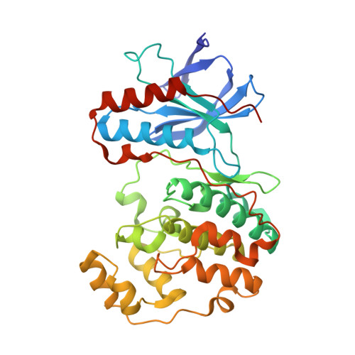
    We describe the structure-guided optimization of the molecular fragments 2-amino-3-benzyloxypyridine 1 (IC(50) 1.3 mM) and 3-(2-(4-pyridyl)ethyl)indole 2 (IC(50) 35 microM) identified using X-ray crystallographic screening of p38alpha MAP kinase. Using two separate case studies, the article focuses on the key compounds synthesized, the structure-activity relationships and the binding mode observations made during this optimization process, resulting in two potent lead series that demonstrate significant increases in activity. We describe the process of compound elaboration either through the growing out from fragments into adjacent pockets or through the conjoining of overlapping fragments and demonstrate that we have exploited the mobile conserved activation loop, consisting in part of Asp168-Phe169-Gly170 (DFG), to generate significant improvements in potency and kinase selectivity.


  • Organizational Affiliation

    Astex Technology, 436 Cambridge Science Park, Milton Road, Cambridge, CB4 0QA, United Kingdom. a.gill@astex-technology.com


Macromolecules
Find similar proteins by:  (by identity cutoff)  |  3D Structure
Entity ID: 1
MoleculeChains Sequence LengthOrganismDetailsImage
MITOGEN-ACTIVATED PROTEIN KINASE 14360Homo sapiensMutation(s): 0 
EC: 2.7.1.37
UniProt & NIH Common Fund Data Resources
Find proteins for Q16539 (Homo sapiens)
Explore Q16539 
Go to UniProtKB:  Q16539
PHAROS:  Q16539
GTEx:  ENSG00000112062 
Entity Groups  
Sequence Clusters30% Identity50% Identity70% Identity90% Identity95% Identity100% Identity
UniProt GroupQ16539
Sequence Annotations
Expand
  • Reference Sequence
Small Molecules
Ligands 1 Unique
IDChains Name / Formula / InChI Key2D Diagram3D Interactions
L11
Query on L11

Download Ideal Coordinates CCD File 
B [auth A]N-[4-CHLORO-3-(PYRIDIN-3-YLOXYMETHYL)-PHENYL]-3-FLUORO-
C23 H25 Cl F N3 O3
YQJVDUKATDECHF-ICSRJNTNSA-N
Binding Affinity Annotations 
IDSourceBinding Affinity
L11 Binding MOAD:  1W83 IC50: 65 (nM) from 1 assay(s)
PDBBind:  1W83 IC50: 65 (nM) from 1 assay(s)
Experimental Data & Validation

Experimental Data

  • Method: X-RAY DIFFRACTION
  • Resolution: 2.50 Å
  • R-Value Free: 0.322 
  • R-Value Work: 0.228 
  • R-Value Observed: 0.233 
  • Space Group: P 21 21 21
Unit Cell:
Length ( Å )Angle ( ˚ )
a = 45.681α = 90
b = 86.101β = 90
c = 126.079γ = 90
Software Package:
Software NamePurpose
REFMACrefinement

Structure Validation

View Full Validation Report


Entry History 

Deposition Data

Revision History  (Full details and data files)

  • Version 1.0: 2005-02-08
    Type: Initial release
  • Version 1.1: 2011-05-08
    Changes: Version format compliance
  • Version 1.2: 2011-07-13
    Changes: Version format compliance
  • Version 1.3: 2018-01-31
    Changes: Database references, Source and taxonomy