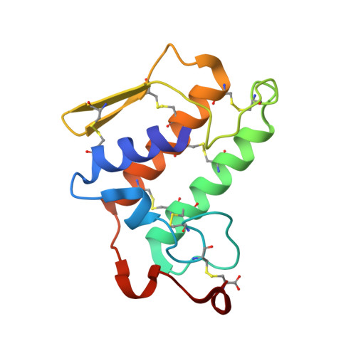
1VKQ

A re-determination of the structure of the triple mutant (K53,56,120M) of phospholipase A2 at 1.6A resolution using sulphur-SAS at 1.54A wavelength


Experimental Data Snapshot

  • Method: X-RAY DIFFRACTION
  • Resolution: 1.60 Å
  • R-Value Free: 0.217 
  • R-Value Work: 0.177 
  • R-Value Observed: 0.179 

wwPDB Validation   3D Report Full Report


This is version 1.6 of the entry. See complete history


Literature

A redetermination of the structure of the triple mutant (K53,56,120M) of phospholipase A2 at 1.6 A resolution using sulfur-SAS at 1.54 A wavelength.

Sekar, K.Rajakannan, V.Velmurugan, D.Yamane, T.Thirumurugan, R.Dauter, M.Dauter, Z.

(2004) Acta Crystallogr D Biol Crystallogr 60: 1586-1590

  • DOI: https://doi.org/10.1107/S090744490401697X
  • Primary Citation of Related Structures:  
    1VKQ

  • PubMed Abstract: 

    The crystal structure of the triple mutant K53,56,120M of bovine pancreatic phospholipase A(2) has been redetermined using sulfur single-wavelength anomalous scattering. The synchrotron data were collected at lambda = 1.54 A and the crystal diffracted to 1.6 A resolution. The program SOLVE was used to locate the heavy atoms and to estimate the initial phases and the resulting map was then subjected to RESOLVE. The output of 455 non-H atoms, including 12 S atoms, one calcium ion and one chloride ion, were then subjected to ARP/wARP followed by REFMAC. With the improved phases, the automatic model building successfully built more than 85% of the 123 residues, excluding the N- and C-terminal residues. The final crystallographic R factor is 17.7% (R(free) = 21.7%). The refined model consists of 954 non-H protein atoms, 165 water O atoms, three 2-methyl-2,4-pentanediol (MPD) molecules, one calcium ion and one chloride ion. The present work is yet another example that shows the utility of single-wavelength anomalous scattering data for solving a protein structure.


  • Organizational Affiliation

    Bioinformatics Centre, Indian Institute of Science, Bangalore 560 012, India. sekar@physics.iisc.ernet.in


Macromolecules
Find similar proteins by:  (by identity cutoff)  |  3D Structure
Entity ID: 1
MoleculeChains Sequence LengthOrganismDetailsImage
Phospholipase A2123Bos taurusMutation(s): 3 
EC: 3.1.1.4
UniProt
Find proteins for P00593 (Bos taurus)
Explore P00593 
Go to UniProtKB:  P00593
Entity Groups  
Sequence Clusters30% Identity50% Identity70% Identity90% Identity95% Identity100% Identity
UniProt GroupP00593
Sequence Annotations
Expand
  • Reference Sequence
Experimental Data & Validation

Experimental Data

  • Method: X-RAY DIFFRACTION
  • Resolution: 1.60 Å
  • R-Value Free: 0.217 
  • R-Value Work: 0.177 
  • R-Value Observed: 0.179 
  • Space Group: P 31 2 1
Unit Cell:
Length ( Å )Angle ( ˚ )
a = 46.05α = 90
b = 46.05β = 90
c = 101.58γ = 120
Software Package:
Software NamePurpose
DENZOdata reduction
REFMACrefinement
SCALEPACKdata scaling

Structure Validation

View Full Validation Report


Entry History 

Deposition Data

Revision History  (Full details and data files)

  • Version 1.0: 2004-08-31
    Type: Initial release
  • Version 1.1: 2008-04-26
    Changes: Version format compliance
  • Version 1.2: 2011-07-13
    Changes: Version format compliance
  • Version 1.3: 2017-10-04
    Changes: Refinement description
  • Version 1.4: 2017-10-11
    Changes: Data collection
  • Version 1.5: 2021-10-27
    Changes: Database references, Derived calculations
  • Version 1.6: 2023-12-27
    Changes: Data collection