1V75

Crystal structure of hemoglobin D from the Aldabra giant tortoise (Geochelone gigantea) at 2.0 A resolution


Experimental Data Snapshot

  • Method: X-RAY DIFFRACTION
  • Resolution: 2.02 Å
  • R-Value Free: 0.233 
  • R-Value Work: 0.188 

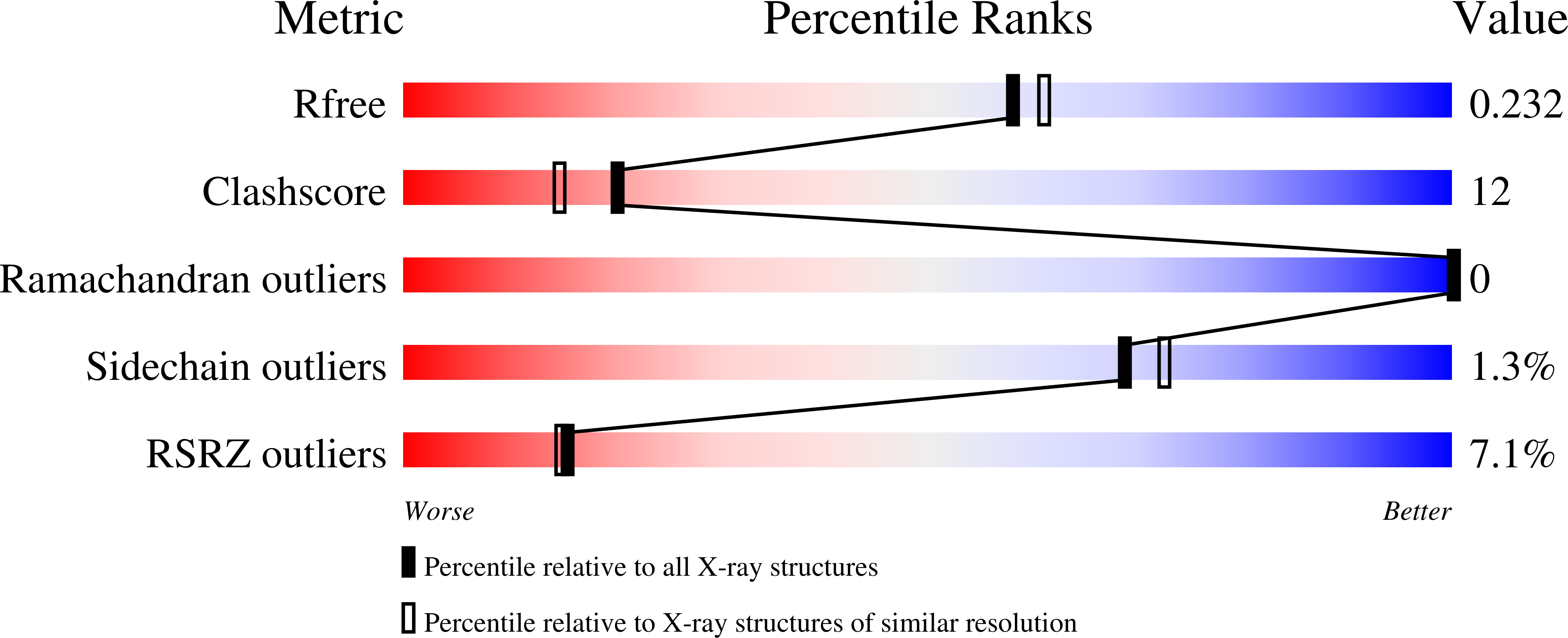
wwPDB Validation   3D Report Full Report


Ligand Structure Quality Assessment 


This is version 1.4 of the entry. See complete history


Literature

Crystallization and preliminary X-ray diffraction study of hemoglobin D from the Aldabra giant tortoise, Geochelone gigantea.

Kuwada, T.Hasegawa, T.Satoh, I.Ishikawa, K.Shishikura, F.

(2003) Protein Pept Lett 10: 422-425

  • DOI: https://doi.org/10.2174/0929866033478799
  • Primary Citation of Related Structures:  
    1V75

  • PubMed Abstract: 

    Hemoglobin D (Hb D) from the Aldabra giant tortoise, Geochelone gigantea, was crystallized by the hanging drop vapor diffusion technique with a precipitant solution containing 10% polyethylene glycol 3350 and 50 mM HEPES-Na, pH 7.5. The Hb D crystals of G. gigantea, which diffract to at least a 2.0 A resolution, belong to the monoclinic space group C2 with unit cell dimensions of a = 112.1 A, b = 62.4 A, c = 54.0 A, and beta = 110.3 degrees. One alphabeta dimer molecule of Hb D existed in an asymmetric unit, with a calculated value of Vm of 2.77 A(3)Da(-1).


  • Organizational Affiliation

    Institute of Quantum Science, Nihon University, Funabashi 274-8501, Japan.


Macromolecules
Find similar proteins by:  (by identity cutoff)  |  3D Structure
Entity ID: 1
MoleculeChains Sequence LengthOrganismDetailsImage
Hemoglobin D alpha chain141Aldabrachelys giganteaMutation(s): 0 
UniProt
Find proteins for P83134 (Aldabrachelys gigantea)
Explore P83134 
Go to UniProtKB:  P83134
Entity Groups  
Sequence Clusters30% Identity50% Identity70% Identity90% Identity95% Identity100% Identity
UniProt GroupP83134
Sequence Annotations
Expand
  • Reference Sequence
Find similar proteins by:  (by identity cutoff)  |  3D Structure
Entity ID: 2
MoleculeChains Sequence LengthOrganismDetailsImage
Hemoglobin A and D beta chain146Aldabrachelys giganteaMutation(s): 0 
UniProt
Find proteins for P83133 (Aldabrachelys gigantea)
Explore P83133 
Go to UniProtKB:  P83133
Entity Groups  
Sequence Clusters30% Identity50% Identity70% Identity90% Identity95% Identity100% Identity
UniProt GroupP83133
Sequence Annotations
Expand
  • Reference Sequence
Small Molecules
Ligands 1 Unique
IDChains Name / Formula / InChI Key2D Diagram3D Interactions
HEM
Query on HEM

Download Ideal Coordinates CCD File 
C [auth A],
D [auth B]
PROTOPORPHYRIN IX CONTAINING FE
C34 H32 Fe N4 O4
KABFMIBPWCXCRK-RGGAHWMASA-L
Experimental Data & Validation

Experimental Data

  • Method: X-RAY DIFFRACTION
  • Resolution: 2.02 Å
  • R-Value Free: 0.233 
  • R-Value Work: 0.188 
  • Space Group: C 1 2 1
Unit Cell:
Length ( Å )Angle ( ˚ )
a = 112.142α = 90
b = 62.374β = 110.26
c = 53.98γ = 90
Software Package:
Software NamePurpose
CNXrefinement
CrystalCleardata reduction
CrystalCleardata scaling
CNXphasing

Structure Validation

View Full Validation Report


Ligand Structure Quality Assessment 


Entry History 

Deposition Data

Revision History  (Full details and data files)

  • Version 1.0: 2003-12-30
    Type: Initial release
  • Version 1.1: 2008-04-27
    Changes: Version format compliance
  • Version 1.2: 2011-07-13
    Changes: Version format compliance
  • Version 1.3: 2019-10-09
    Changes: Data collection, Database references
  • Version 1.4: 2023-12-27
    Changes: Data collection, Database references, Derived calculations