1UW1

A Novel ADP- and Zinc-binding fold from function-directed in vitro evolution


Experimental Data Snapshot

  • Method: X-RAY DIFFRACTION
  • Resolution: 1.94 Å
  • R-Value Free: 0.228 
  • R-Value Work: 0.200 

wwPDB Validation   3D Report Full Report


Ligand Structure Quality Assessment 


This is version 1.2 of the entry. See complete history


Literature

A Novel Adp- and Zinc-Binding Fold from Function-Directed in Vitro Evolution

Lo Surdo, P.Walsh, M.A.Sollazzo, M.

(2004) Nat Struct Mol Biol 11: 382

  • DOI: https://doi.org/10.1038/nsmb745
  • Primary Citation of Related Structures:  
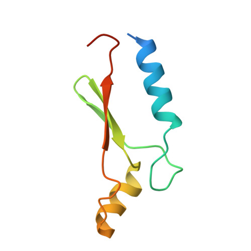
    1UW1

  • PubMed Abstract: 

    A great challenge to biologists is to create proteins with novel folds and tailored functions. As an alternative to de novo protein design, we investigated the structure of a randomly generated protein targeted to bind ATP. The crystal structure reveals a novel alpha/beta fold bound to its ligand, representing both the first protein structure derived from in vitro evolution and the first nucleotide-binding protein stabilized by a zinc ion.


  • Organizational Affiliation

    Istituto di Ricerche di Biologia Molecolare (IRBM), Via Pontina Km 30.600, 00040 Pomezia, Roma, Italy. paola_losurdo@merck.com


Macromolecules
Find similar proteins by:  (by identity cutoff)  |  3D Structure
Entity ID: 1
MoleculeChains Sequence LengthOrganismDetailsImage
ARTIFICIAL NUCLEOTIDE BINDING PROTEIN (ANBP)80synthetic constructMutation(s): 0 
Entity Groups  
Sequence Clusters30% Identity50% Identity70% Identity90% Identity95% Identity100% Identity
Sequence Annotations
Expand
  • Reference Sequence
Small Molecules
Ligands 2 Unique
IDChains Name / Formula / InChI Key2D Diagram3D Interactions
ADP
Query on ADP

Download Ideal Coordinates CCD File 
B [auth A]ADENOSINE-5'-DIPHOSPHATE
C10 H15 N5 O10 P2
XTWYTFMLZFPYCI-KQYNXXCUSA-N
ZN
Query on ZN

Download Ideal Coordinates CCD File 
C [auth A]ZINC ION
Zn
PTFCDOFLOPIGGS-UHFFFAOYSA-N
Binding Affinity Annotations 
IDSourceBinding Affinity
ADP Binding MOAD:  1UW1 Kd: 100 (nM) from 1 assay(s)
Experimental Data & Validation

Experimental Data

  • Method: X-RAY DIFFRACTION
  • Resolution: 1.94 Å
  • R-Value Free: 0.228 
  • R-Value Work: 0.200 
  • Space Group: P 32 2 1
Unit Cell:
Length ( Å )Angle ( ˚ )
a = 71.077α = 90
b = 71.077β = 90
c = 54.883γ = 120
Software Package:
Software NamePurpose
REFMACrefinement
DENZOdata reduction
SCALEPACKdata scaling
SHELXDphasing

Structure Validation

View Full Validation Report


Ligand Structure Quality Assessment 


Entry History 

Deposition Data

Revision History  (Full details and data files)

  • Version 1.0: 2004-03-26
    Type: Initial release
  • Version 1.1: 2015-10-21
    Changes: Derived calculations, Non-polymer description, Other, Source and taxonomy, Structure summary, Version format compliance
  • Version 1.2: 2019-05-15
    Changes: Data collection, Experimental preparation, Other