The Crystal Structure of the Periplasmic Domain of the Type II Secretion System Protein Epsm from Vibrio Cholerae: The Simplest Version of the Ferredoxin Fold
Abendroth, J., Rice, A., Mcluskey, K., Bagdasarian, M., Hol, W.G.J.(2004) J Mol Biol 338: 585
- PubMed: 15081815
- DOI: https://doi.org/10.1016/j.jmb.2004.01.064
- Primary Citation of Related Structures:
1UV7 - PubMed Abstract:
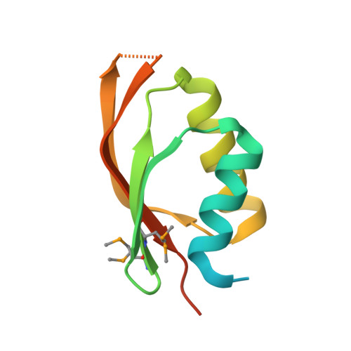
The terminal branch of the general secretion pathway (Gsp or type II secretion system) is used by several pathogenic bacteria for the secretion of their virulence factors across the outer membrane. In these secretion systems, a complex of 12-15 Gsp proteins spans from the pore in the outer membrane via several associated signal or energy-transducing proteins in the inner membrane to a regulating ATPase in the cytosol. The human pathogen Vibrio cholerae uses such a system, called the Eps system, for the export of the cholera toxin and other virulence factors from its periplasm into the lumen of the gastrointestinal tract of the host. Here, we report the atomic structure of the periplasmic domain of the EpsM protein from V.cholerae, which is a part of the interface between the regulating part and the rest of the Eps system. The crystal structure was determined by Se-Met MAD phasing and the model was refined to 1.7A resolution. The monomer consists of two alphabetabeta-subdomains forming a sandwich of two alpha-helices and a four-stranded antiparallel beta-sheet. In the dimer, a deep cleft with a polar rim and a hydrophobic bottom made by conserved residues is located between the monomers. This cleft contains an extra electron density suggesting that this region might serve as a binding site of an unknown ligand or part of a protein partner. Unexpectedly, the fold of the periplasmic domain of EpsM is an undescribed circular permutation of the ferredoxin fold.
Organizational Affiliation:
Department of Biochemistry, Biomolecular Structure Center, School of Medicine, University of Washington, Box 357742, Seattle, WA 98195-7242, USA.