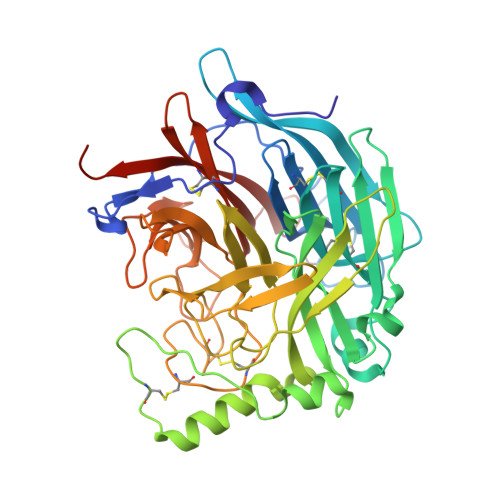
Second Sialic Acid Binding Site in Newcastle Disease Virus Hemagglutinin-Neuraminidase: Implications for Fusion
Zaitsev, V., Von Itzstein, M., Groves, D., Kiefel, M., Takimoto, T., Portner, A., Taylor, G.(2004) J Virol 78: 3733
- PubMed: 15016893
- DOI: https://doi.org/10.1128/jvi.78.7.3733-3741.2004
- Primary Citation of Related Structures:
1USR, 1USX - PubMed Abstract:
Paramyxoviruses are the leading cause of respiratory disease in children. Several paramyxoviruses possess a surface glycoprotein, the hemagglutinin-neuraminidase (HN), that is involved in attachment to sialic acid receptors, promotion of fusion, and removal of sialic acid from infected cells and progeny virions. Previously we showed that Newcastle disease virus (NDV) HN contained a pliable sialic acid recognition site that could take two states, a binding state and a catalytic state. Here we present evidence for a second sialic acid binding site at the dimer interface of HN and present a model for its involvement in cell fusion. Three different crystal forms of NDV HN now reveal identical tetrameric arrangements of HN monomers, perhaps indicative of the tetramer association found on the viral surface.
Organizational Affiliation:
Centre for Biomolecular Sciences, University of St. Andrews, St. Andrews, Fife KY16 9ST, United Kingdom.