1URH

The "Rhodanese" fold and catalytic mechanism of 3-mercaptopyruvate sulfotransferases: Crystal structure of SseA from Escherichia coli


Experimental Data Snapshot

  • Method: X-RAY DIFFRACTION
  • Resolution: 2.80 Å
  • R-Value Free: 0.289 
  • R-Value Work: 0.234 
  • R-Value Observed: 0.234 

wwPDB Validation   3D Report Full Report


This is version 1.3 of the entry. See complete history


Literature

The "Rhodanese" Fold and Catalytic Mechanism of 3-Mercaptopyruvate Sulfurtransferases: Crystal Structure of Ssea from Escherichia Coli

Spallarossa, A.Forlani, F.Carpen, A.Armirotti, A.Pagani, S.Bolognesi, M.Bordo, D.

(2004) J Mol Biol 335: 583

  • DOI: https://doi.org/10.1016/j.jmb.2003.10.072
  • Primary Citation of Related Structures:  
    1URH

  • PubMed Abstract: 

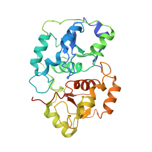
    3-Mercaptopyruvate sulfurtransferases (MSTs) catalyze, in vitro, the transfer of a sulfur atom from substrate to cyanide, yielding pyruvate and thiocyanate as products. They display clear structural homology with the protein fold observed in the rhodanese sulfurtransferase family, composed of two structurally related domains. The role of MSTs in vivo, as well as their detailed molecular mechanisms of action have been little investigated. Here, we report the crystal structure of SseA, a MST from Escherichia coli, which is the first MST three-dimensional structure disclosed to date. SseA displays specific structural differences relative to eukaryotic and prokaryotic rhodaneses. In particular, conformational variation of the rhodanese active site loop, hosting the family invariant catalytic Cys residue, may support a new sulfur transfer mechanism involving Cys237 as the nucleophilic species and His66, Arg102 and Asp262 as residues assisting catalysis.


  • Organizational Affiliation

    Dipartimento di Fisica-INFM, Centro di Eccellenza per la Ricerca Biomedica, Universita' di Genova; Via Dodecaneso 33, 16146 Genoa, Italy.


Macromolecules
Find similar proteins by:  (by identity cutoff)  |  3D Structure
Entity ID: 1
MoleculeChains Sequence LengthOrganismDetailsImage
3-MERCAPTOPYRUVATE SULFURTRANSFERASE
A, B
280Escherichia coliMutation(s): 0 
EC: 2.8.1.2
UniProt
Find proteins for P31142 (Escherichia coli (strain K12))
Explore P31142 
Go to UniProtKB:  P31142
Entity Groups  
Sequence Clusters30% Identity50% Identity70% Identity90% Identity95% Identity100% Identity
UniProt GroupP31142
Sequence Annotations
Expand
  • Reference Sequence
Small Molecules
Ligands 1 Unique
IDChains Name / Formula / InChI Key2D Diagram3D Interactions
SO3
Query on SO3

Download Ideal Coordinates CCD File 
C [auth B]SULFITE ION
O3 S
LSNNMFCWUKXFEE-UHFFFAOYSA-L
Modified Residues  1 Unique
IDChains TypeFormula2D DiagramParent
CSS
Query on CSS
A, B
L-PEPTIDE LINKINGC3 H7 N O2 S2CYS
Experimental Data & Validation

Experimental Data

  • Method: X-RAY DIFFRACTION
  • Resolution: 2.80 Å
  • R-Value Free: 0.289 
  • R-Value Work: 0.234 
  • R-Value Observed: 0.234 
  • Space Group: P 43
Unit Cell:
Length ( Å )Angle ( ˚ )
a = 150.17α = 90
b = 150.17β = 90
c = 37.93γ = 90
Software Package:
Software NamePurpose
CNSrefinement
DENZOdata reduction
SCALEPACKdata scaling
AMoREphasing

Structure Validation

View Full Validation Report


Entry History 

Deposition Data

Revision History  (Full details and data files)

  • Version 1.0: 2003-12-18
    Type: Initial release
  • Version 1.1: 2011-05-08
    Changes: Version format compliance
  • Version 1.2: 2011-07-13
    Changes: Version format compliance
  • Version 1.3: 2023-12-13
    Changes: Data collection, Database references, Derived calculations, Other, Refinement description