


Crystal Structure of Ydhf the E.Coli Aldo-Keto Reductase Ydhf
Jeudy, S., Claverie, J.M., Abergel, C.To be published.
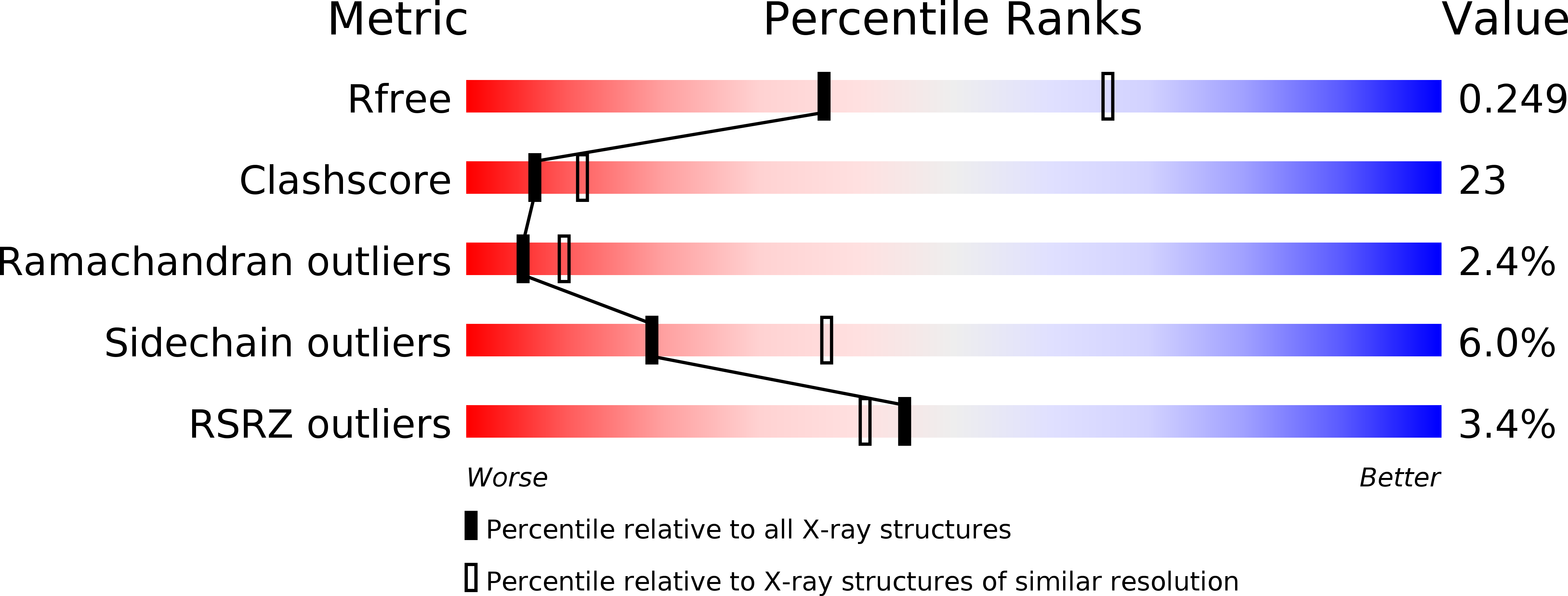
Experimental Data Snapshot
wwPDB Validation 3D Report Full Report
Entity ID: 1 | |||||
|---|---|---|---|---|---|
| Molecule | Chains | Sequence Length | Organism | Details | Image |
| HYPOTHETICAL OXIDOREDUCTASE YDHF | A [auth M] | 319 | Escherichia coli K-12 | Mutation(s): 0 EC: 1 | 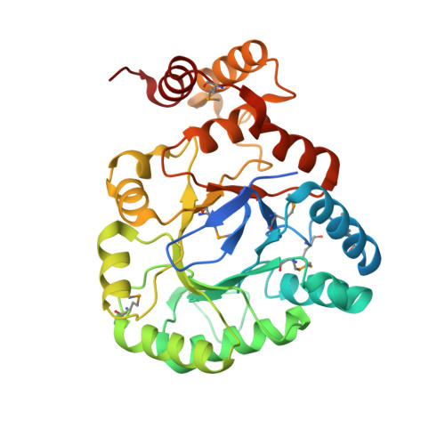 |
UniProt | |||||
Find proteins for P76187 (Escherichia coli (strain K12)) Explore P76187 Go to UniProtKB: P76187 | |||||
Entity Groups | |||||
| Sequence Clusters | 30% Identity50% Identity70% Identity90% Identity95% Identity100% Identity | ||||
| UniProt Group | P76187 | ||||
Sequence AnnotationsExpand | |||||
| |||||
| Ligands 1 Unique | |||||
|---|---|---|---|---|---|
| ID | Chains | Name / Formula / InChI Key | 2D Diagram | 3D Interactions | |
| SO4 Query on SO4 | B [auth M] | SULFATE ION O4 S QAOWNCQODCNURD-UHFFFAOYSA-L |  | ||
| Modified Residues 1 Unique | |||||
|---|---|---|---|---|---|
| ID | Chains | Type | Formula | 2D Diagram | Parent |
| MSE Query on MSE | A [auth M] | L-PEPTIDE LINKING | C5 H11 N O2 Se |  | MET |
| Length ( Å ) | Angle ( ˚ ) |
|---|---|
| a = 87.722 | α = 90 |
| b = 87.722 | β = 90 |
| c = 66.168 | γ = 120 |
| Software Name | Purpose |
|---|---|
| CNS | refinement |
| MOSFLM | data reduction |
| SCALA | data scaling |
| autoSHARP | phasing |
RCSB PDB (citation) is hosted by
RCSB PDB is a member of the

