1UCR

Three-dimensional crystal structure of dissimilatory sulfite reductase D (DsrD)


Experimental Data Snapshot

  • Method: X-RAY DIFFRACTION
  • Resolution: 1.20 Å
  • R-Value Free: 0.180 
  • R-Value Work: 0.136 
  • R-Value Observed: 0.138 

wwPDB Validation   3D Report Full Report


This is version 1.3 of the entry. See complete history


Literature

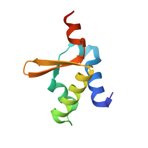
Crystal Structure of Dissimilatory Sulfite Reductase D (DsrD) Protein-Possible Interaction with B- and Z-DNA by Its Winged-Helix Motif

Mizuno, N.Voordouw, G.Miki, K.Sarai, A.Higuchi, Y.

(2003) Structure 11: 1133-1140

  • DOI: https://doi.org/10.1016/s0969-2126(03)00156-4
  • Primary Citation of Related Structures:  
    1UCR

  • PubMed Abstract: 

    The crystal structure of DsrD from Desulfovibrio vulgaris Hildenborough has been determined at 1.2 A resolution. DsrD is in a dimeric form in the crystal, and five sulfate anions were located on the surface. The structure of DsrD comprises a winged-helix motif, which shows the highest structural homology to similar motifs found in Z-DNA binding proteins and some B-DNA binding proteins. The core structure of the molecule is constructed by intramolecular interactions of hydrophobic residues, which are well conserved in DNA binding proteins, suggesting that these proteins belong to the same superfamily on the basis of the structure. These results indicate a possible role of DsrD in transcription or translation of genes for enzymes catalyzing dissimilatory sulfite reduction.


  • Organizational Affiliation

    Division of Chemistry, Graduate School of Science, Kyoto University, Sakyo, Kyoto 606-8502, Japan.


Macromolecules
Find similar proteins by:  (by identity cutoff)  |  3D Structure
Entity ID: 1
MoleculeChains Sequence LengthOrganismDetailsImage
Protein dsvD
A, B
78Nitratidesulfovibrio vulgarisMutation(s): 0 
UniProt
Find proteins for Q46582 (Desulfovibrio vulgaris (strain ATCC 29579 / DSM 644 / NCIMB 8303 / VKM B-1760 / Hildenborough))
Explore Q46582 
Go to UniProtKB:  Q46582
Entity Groups  
Sequence Clusters30% Identity50% Identity70% Identity90% Identity95% Identity100% Identity
UniProt GroupQ46582
Sequence Annotations
Expand
  • Reference Sequence
Experimental Data & Validation

Experimental Data

  • Method: X-RAY DIFFRACTION
  • Resolution: 1.20 Å
  • R-Value Free: 0.180 
  • R-Value Work: 0.136 
  • R-Value Observed: 0.138 
  • Space Group: P 21 21 21
Unit Cell:
Length ( Å )Angle ( ˚ )
a = 60.025α = 90
b = 64.559β = 90
c = 45.218γ = 90
Software Package:
Software NamePurpose
SHELXmodel building
SHELXL-97refinement
MOSFLMdata reduction
CCP4data scaling
MLPHAREphasing

Structure Validation

View Full Validation Report


Entry History 

Deposition Data

Revision History  (Full details and data files)

  • Version 1.0: 2003-10-14
    Type: Initial release
  • Version 1.1: 2008-04-27
    Changes: Version format compliance
  • Version 1.2: 2011-07-13
    Changes: Version format compliance
  • Version 1.3: 2023-12-27
    Changes: Data collection, Database references, Derived calculations