1TXX

ACTIVE-SITE VARIANT OF E.COLI THIOREDOXIN


Experimental Data Snapshot

  • Method: X-RAY DIFFRACTION
  • Resolution: 2.20 Å
  • R-Value Work: 0.171 

wwPDB Validation   3D Report Full Report


This is version 1.5 of the entry. See complete history


Literature

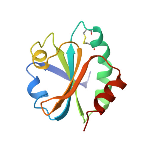
The CXXC motif: crystal structure of an active-site variant of Escherichia coli thioredoxin.

Schultz, L.W.Chivers, P.T.Raines, R.T.

(1999) Acta Crystallogr D Biol Crystallogr 55: 1533-1538

  • DOI: https://doi.org/10.1107/s0907444999008756
  • Primary Citation of Related Structures:  
    1TXX

  • PubMed Abstract: 

    The 2.2 A crystalline structure of an oxidized active-site variant of Escherichia coli thioredoxin (Trx) has been solved. Trx is a 12 kDa enzyme which catalyzes the oxidation of dithiols and the reduction and isomerization of disulfides in other proteins. Its active site contains the common structural motif CXXC. Protein-disulfide isomerase (PDI), a 57 kDa homolog of Trx, contains four Trx-like domains. The three-dimensional structure of PDI is unknown. PDI-deficient Saccharomyces cerevisiae are inviable. An active-site variant of Trx which complements PDI-deficient yeast has the active-site sequence Cys32-Val33-Trp34-Cys35 (CVWC). The reduction potential of oxidized CVWC Trx (E degrees ' = -0.230 V) is altered significantly from that of the wild-type enzyme (E degrees ' = -0.270 V). However, the structure of the oxidized CVWC enzyme is almost identical to that of wild-type Trx. The addition of valine and tryptophan in the active site is likely to increase the reduction potential, largely by decreasing the pK(a) of the Cys32 thiol in the reduced enzyme. Unlike in wild-type Trx, significant protein-protein contacts occur in the crystal. Protein molecules related by a crystallographic twofold axis form a dimer in the crystal. The dimer forms as an extension of the twisted mixed beta-sheet which composes the backbone of each Trx structure.


  • Organizational Affiliation

    Department of Biochemistry, University of Wisconsin-Madison, Madison, WI 53706, USA.


Macromolecules
Find similar proteins by:  (by identity cutoff)  |  3D Structure
Entity ID: 1
MoleculeChains Sequence LengthOrganismDetailsImage
PROTEIN (THIOREDOXIN)108Escherichia coliMutation(s): 2 
EC: 1.8
UniProt
Find proteins for P0AA25 (Escherichia coli (strain K12))
Explore P0AA25 
Go to UniProtKB:  P0AA25
Entity Groups  
Sequence Clusters30% Identity50% Identity70% Identity90% Identity95% Identity100% Identity
UniProt GroupP0AA25
Sequence Annotations
Expand
  • Reference Sequence
Small Molecules
Ligands 1 Unique
IDChains Name / Formula / InChI Key2D Diagram3D Interactions
CU
Query on CU

Download Ideal Coordinates CCD File 
B [auth A]COPPER (II) ION
Cu
JPVYNHNXODAKFH-UHFFFAOYSA-N
Experimental Data & Validation

Experimental Data

  • Method: X-RAY DIFFRACTION
  • Resolution: 2.20 Å
  • R-Value Work: 0.171 
  • Space Group: P 43 21 2
Unit Cell:
Length ( Å )Angle ( ˚ )
a = 42.53α = 90
b = 42.53β = 90
c = 105.24γ = 90
Software Package:
Software NamePurpose
XDSdata scaling
XSCALIBREdata reduction
AMoREphasing
TNTrefinement
XDSdata reduction
XSCALIBREdata scaling

Structure Validation

View Full Validation Report


Entry History 

Deposition Data

Revision History  (Full details and data files)

  • Version 1.0: 1999-09-13
    Type: Initial release
  • Version 1.1: 2008-04-26
    Changes: Version format compliance
  • Version 1.2: 2011-07-13
    Changes: Version format compliance
  • Version 1.3: 2017-10-04
    Changes: Advisory, Refinement description
  • Version 1.4: 2018-03-14
    Changes: Database references
  • Version 1.5: 2023-08-23
    Changes: Advisory, Data collection, Database references, Derived calculations, Refinement description