1TWQ

Crystal structure of the C-terminal PGN-binding domain of human PGRP-Ialpha in complex with PGN analog muramyl tripeptide


Experimental Data Snapshot

  • Method: X-RAY DIFFRACTION
  • Resolution: 2.30 Å
  • R-Value Free: 0.252 
  • R-Value Work: 0.222 
  • R-Value Observed: 0.222 

wwPDB Validation   3D Report Full Report


Ligand Structure Quality Assessment 


This is version 3.0 of the entry. See complete history


Literature

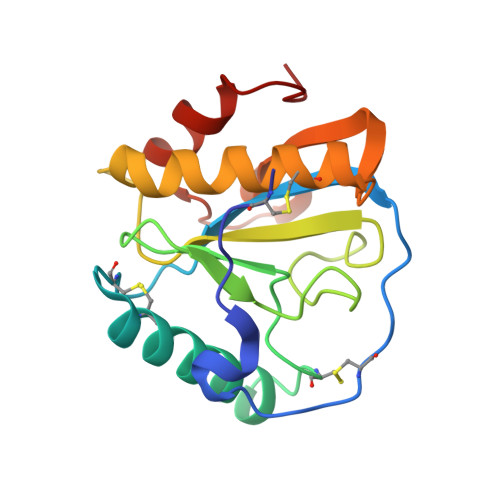
Structural basis for peptidoglycan binding by peptidoglycan recognition proteins

Guan, R.Roychowdhury, A.Ember, B.Kumar, S.Boons, G.-A.Mariuzza, R.A.

(2004) Proc Natl Acad Sci U S A 101: 17168-17173

  • DOI: https://doi.org/10.1073/pnas.0407856101
  • Primary Citation of Related Structures:  
    1TWQ

  • PubMed Abstract: 

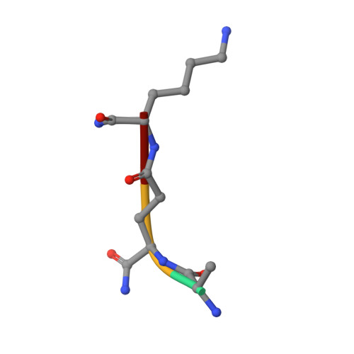
    Peptidoglycan (PGN) recognition proteins (PGRPs) are pattern-recognition receptors of the innate immune system that bind and, in some cases, hydrolyze bacterial PGNs. We determined the crystal structure, at 2.30-A resolution, of the C-terminal PGN-binding domain of human PGRP-Ialpha in complex with a muramyl tripeptide representing the core of lysine-type PGNs from Gram-positive bacteria. The peptide stem of the ligand is buried at the deep end of a long binding groove, with N-acetylmuramic acid situated in the middle of the groove, whose shallow end can accommodate a linked N-acetylglucosamine. Although most interactions are with the peptide, the glycan moiety also seems to be essential for specific recognition by PGRPs. Conservation of key PGN-contacting residues shows that all PGRPs employ this basic PGN-binding mode. The structure pinpoints variable residues that likely mediate discrimination between lysine- and diaminopimelic acid-type PGNs. We also propose a mechanism for PGN hydrolysis by Zn(2+)-containing PGRPs.


  • Organizational Affiliation

    Center for Advanced Research in Biotechnology, W. M. Keck Laboratory for Structural Biology, University of Maryland Biotechnology Institute, Rockville, MD 20850, USA.


Macromolecules
Find similar proteins by:  (by identity cutoff)  |  3D Structure
Entity ID: 1
MoleculeChains Sequence LengthOrganismDetailsImage
peptidoglycan recognition protein-I-alpha165Homo sapiensMutation(s): 0 
Gene Names: PGLYRP3PGRPIA
UniProt & NIH Common Fund Data Resources
Find proteins for Q96LB9 (Homo sapiens)
Explore Q96LB9 
Go to UniProtKB:  Q96LB9
PHAROS:  Q96LB9
GTEx:  ENSG00000159527 
Entity Groups  
Sequence Clusters30% Identity50% Identity70% Identity90% Identity95% Identity100% Identity
UniProt GroupQ96LB9
Sequence Annotations
Expand
  • Reference Sequence

Find similar proteins by:  Sequence   |   3D Structure  

Entity ID: 2
MoleculeChains Sequence LengthOrganismDetailsImage
muramyl tripeptideB [auth P]4synthetic constructMutation(s): 0 
Sequence Annotations
Expand
  • Reference Sequence
Small Molecules
Ligands 2 Unique
IDChains Name / Formula / InChI Key2D Diagram3D Interactions
AMU
Query on AMU

Download Ideal Coordinates CCD File 
D [auth P]N-acetyl-beta-muramic acid
C11 H19 N O8
MNLRQHMNZILYPY-YVNCZSHWSA-N
NI
Query on NI

Download Ideal Coordinates CCD File 
C [auth A]NICKEL (II) ION
Ni
VEQPNABPJHWNSG-UHFFFAOYSA-N
Modified Residues  1 Unique
IDChains TypeFormula2D DiagramParent
GMA
Query on GMA
B [auth P]L-PEPTIDE LINKINGC5 H10 N2 O3GLU
Experimental Data & Validation

Experimental Data

  • Method: X-RAY DIFFRACTION
  • Resolution: 2.30 Å
  • R-Value Free: 0.252 
  • R-Value Work: 0.222 
  • R-Value Observed: 0.222 
  • Space Group: P 32 2 1
Unit Cell:
Length ( Å )Angle ( ˚ )
a = 104.511α = 90
b = 104.511β = 90
c = 42.05γ = 120
Software Package:
Software NamePurpose
CrystalCleardata collection
CrystalCleardata reduction
AMoREphasing
CNSrefinement
CrystalCleardata scaling

Structure Validation

View Full Validation Report


Ligand Structure Quality Assessment 


Entry History 

Deposition Data

Revision History  (Full details and data files)

  • Version 1.0: 2004-12-14
    Type: Initial release
  • Version 1.1: 2008-04-30
    Changes: Version format compliance
  • Version 1.2: 2011-07-13
    Changes: Version format compliance
  • Version 1.3: 2011-11-16
    Changes: Atomic model
  • Version 1.4: 2017-10-11
    Changes: Advisory, Refinement description
  • Version 1.5: 2020-07-29
    Type: Remediation
    Reason: Carbohydrate remediation
    Changes: Advisory, Data collection, Derived calculations, Structure summary
  • Version 1.6: 2023-08-23
    Changes: Data collection, Database references, Refinement description, Structure summary
  • Version 2.0: 2023-11-15
    Changes: Atomic model, Data collection, Derived calculations
  • Version 3.0: 2024-02-28
    Changes: Advisory, Atomic model, Data collection, Database references, Derived calculations, Polymer sequence, Source and taxonomy, Structure summary