1TTV

NMR Structure of a Complex Between MDM2 and a Small Molecule Inhibitor


Experimental Data Snapshot

  • Method: SOLUTION NMR
  • Conformers Calculated: 42 
  • Conformers Submitted: 18 
  • Selection Criteria: structures with acceptable covalent geometry,structures with the least restraint violations 

wwPDB Validation   3D Report Full Report


This is version 1.3 of the entry. See complete history


Literature

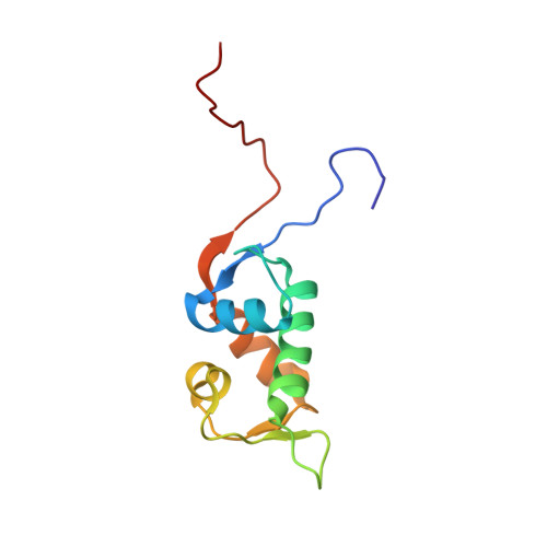
NMR structure of a complex between MDM2 and a small molecule inhibitor.

Fry, D.C.Emerson, S.D.Palme, S.Vu, B.T.Liu, C.M.Podlaski, F.

(2004) J Biomol NMR 30: 163-173

  • DOI: https://doi.org/10.1023/B:JNMR.0000048856.84603.9b
  • Primary Citation of Related Structures:  
    1TTV

  • PubMed Abstract: 

    MDM2 is a regulator of cell growth processes that acts by binding to the tumor suppressor protein p53 and ultimately restraining its activity. While inactivation of p53 by mutation is commonly observed in human cancers, a substantial percentage of tumors express wild type p53. In many of these cases, MDM2 is overexpressed, and it is believed that suppression of MDM2 activity could yield therapeutic benefits. Therefore, we have been focusing on the p53-MDM2 interaction as the basis of a drug discovery program and have been able to develop a series of small molecule inhibitors. We herein report a high resolution NMR structure of a complex between the p53-binding domain of MDM2 and one of these inhibitors. The form of MDM2 utilized was an engineered hybrid between the human and Xenopus sequences, which provided a favorable combination of relevancy and stability. The inhibitor is found to bind in the same site as does a highly potent peptide fragment of p53. The inhibitor is able to successfully mimic the peptide by duplicating interactions in three subpockets normally made by amino acid sidechains, and by utilizing a scaffold that presents substituents with rigidity and spatial orientation comparable to that provided by the alpha helical backbone of the peptide. The structure also suggests opportunities for modifying the inhibitor to increase its potency.


  • Organizational Affiliation

    Roche Research Center, Hoffmann-La Roche, Inc. 340 Kingsland Street, Nutley, NJ 07110, USA. david.fry@roche.com


Macromolecules
Find similar proteins by:  (by identity cutoff)  |  3D Structure
Entity ID: 1
MoleculeChains Sequence LengthOrganismDetailsImage
Ubiquitin-protein ligase E3 Mdm2107Xenopus laevisMutation(s): 3 
Gene Names: MDM2
EC: 6.3.2
UniProt
Find proteins for P56273 (Xenopus laevis)
Explore P56273 
Go to UniProtKB:  P56273
Entity Groups  
Sequence Clusters30% Identity50% Identity70% Identity90% Identity95% Identity100% Identity
UniProt GroupP56273
Sequence Annotations
Expand
  • Reference Sequence
Small Molecules
Ligands 1 Unique
IDChains Name / Formula / InChI Key2D Diagram3D Interactions
IMY
Query on IMY

Download Ideal Coordinates CCD File 
B [auth A]1-{[4,5-BIS(4-CHLOROPHENYL)-2-(2-ISOPROPOXY-4-METHOXYPHENYL)-4,5-DIHYDRO-1H-IMIDAZOL-1-YL]CARBONYL}PIPERAZINE
C30 H32 Cl2 N4 O3
ZXIPEZDMQNYFOO-WUFINQPMSA-N
Binding Affinity Annotations 
IDSourceBinding Affinity
IMY PDBBind:  1TTV IC50: 160 (nM) from 1 assay(s)
Experimental Data & Validation

Experimental Data

  • Method: SOLUTION NMR
  • Conformers Calculated: 42 
  • Conformers Submitted: 18 
  • Selection Criteria: structures with acceptable covalent geometry,structures with the least restraint violations 

Structure Validation

View Full Validation Report


Entry History 

Deposition Data

Revision History  (Full details and data files)

  • Version 1.0: 2005-01-04
    Type: Initial release
  • Version 1.1: 2008-04-30
    Changes: Version format compliance
  • Version 1.2: 2011-07-13
    Changes: Version format compliance
  • Version 1.3: 2021-10-27
    Changes: Data collection, Database references, Derived calculations