


The Geometry of the Reactive Site and of the Peptide Groups in Trypsin, Trypsinogen and its Complexes with Inhibitors
Marquart, M., Walter, J., Deisenhofer, J., Bode, W., Huber, R.(1983) Acta Crystallogr B 39: 480
Experimental Data Snapshot
(1983) Acta Crystallogr B 39: 480
Entity ID: 1 | |||||
|---|---|---|---|---|---|
| Molecule | Chains | Sequence Length | Organism | Details | Image |
| BETA-TRYPSIN | 223 | Bos taurus | Mutation(s): 0 EC: 3.4.21.4 | 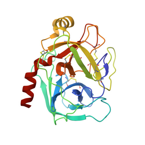 | |
UniProt | |||||
Find proteins for P00760 (Bos taurus) Explore P00760 Go to UniProtKB: P00760 | |||||
Entity Groups | |||||
| Sequence Clusters | 30% Identity50% Identity70% Identity90% Identity95% Identity100% Identity | ||||
| UniProt Group | P00760 | ||||
Sequence AnnotationsExpand | |||||
| |||||
| Ligands 3 Unique | |||||
|---|---|---|---|---|---|
| ID | Chains | Name / Formula / InChI Key | 2D Diagram | 3D Interactions | |
| APA Query on APA | D [auth A] | (2S)-3-(4-carbamimidoylphenyl)-2-hydroxypropanoic acid C10 H12 N2 O3 FAFAPKBWTCDDJC-QMMMGPOBSA-N |  | ||
| SO4 Query on SO4 | B [auth A] | SULFATE ION O4 S QAOWNCQODCNURD-UHFFFAOYSA-L |  | ||
| CA Query on CA | C [auth A] | CALCIUM ION Ca BHPQYMZQTOCNFJ-UHFFFAOYSA-N |  | ||
| Length ( Å ) | Angle ( ˚ ) |
|---|---|
| a = 54.9 | α = 90 |
| b = 58.5 | β = 90 |
| c = 67.8 | γ = 90 |
RCSB PDB (citation) is hosted by
RCSB PDB is a member of the

