Crystal Structure of the Disintegrin Heterodimer from Saw-Scaled Viper (Echis carinatus) at 1.9 A Resolution
Bilgrami, S., Yadav, S., Kaur, P., Sharma, S., Perbandt, M., Betzel, C., Singh, T.P.(2005) Biochemistry 44: 11058-11066
- PubMed: 16101289
- DOI: https://doi.org/10.1021/bi050849y
- Primary Citation of Related Structures:
1TEJ - PubMed Abstract:
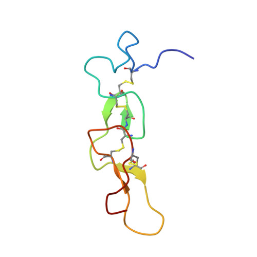
Disintegrins constitute a family of potent polypeptide inhibitors of integrins. Integrins are transmembrane heterodimeric molecules involved in cell-cell and cell-extracellular matrix interactions. They are involved in many diseases such as cancer and thrombosis. Thus, disintegrins have a great potential as anticancer and antithrombotic agents. A novel heterodimeric disintegrin was isolated from the venom of saw-scaled viper (Echis carinatus) and was crystallized. The crystals diffracted to 1.9 A resolution and belonged to space group P4(3)2(1)2. The data indicated the presence of a pseudosymmetry. The structure was solved by applying origin shifts to the disintegrin homodimer schistatin solved in space group I4(1)22 with similar cell dimensions. The structure refined to the final R(cryst)/R(free) factors of 0.213/0.253. The notable differences are observed between the loops, (Gln39-Asp48) containing the important Arg42-Gly43-Asp44, of the present heterodimer and schistatin. These differences are presumably due to the presence of two glycines at positions 43 and 46 that allow the molecule to adopt variable conformations. A comparative analysis of the surface-charge distributions of various disintegrins showed that the charge distribution on monomeric disintegrins occurred uniformly over the whole surface of the molecule, while in the dimeric disintegrins, the charge is distributed only on one face. Such a feature may be important in the binding of two integrins to a single dimeric disintegrin. The phylogenetic analysis developed on the basis of amino acid sequence and three-dimensional structures indicates that the protein diversification and evolution presumably took place from the medium disintegrins and both the dimeric and short disintegrins evolved from them.
Organizational Affiliation:
Department of Biophysics, All India Institute of Medical Sciences, New Delhi 110 029, India.