1TEI

STRUCTURE OF CONCANAVALIN A COMPLEXED TO BETA-D-GLCNAC (1,2)ALPHA-D-MAN-(1,6)[BETA-D-GLCNAC(1,2)ALPHA-D-MAN (1,6)]ALPHA-D-MAN


Experimental Data Snapshot

  • Method: X-RAY DIFFRACTION
  • Resolution: 2.70 Å
  • R-Value Free: 0.219 
  • R-Value Work: 0.179 
  • R-Value Observed: 0.179 

wwPDB Validation   3D Report Full Report


This is version 2.1 of the entry. See complete history


Literature

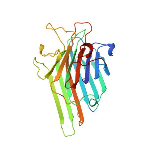
Concanavalin A distorts the beta-GlcNAc-(1-->2)-Man linkage of beta-GlcNAc-(1-->2)-alpha-Man-(1-->3)-[beta-GlcNAc-(1-->2)-alpha-Man- (1-->6)]-Man upon binding.

Moothoo, D.N.Naismith, J.H.

(1998) Glycobiology 8: 173-181

  • DOI: https://doi.org/10.1093/glycob/8.2.173
  • Primary Citation of Related Structures:  
    1TEI

  • PubMed Abstract: 

    Carbohydrate recognition by proteins is a key event in many biological processes. Concanavalin A is known to specifically recognize the pentasaccharide core (beta-GlcNAc-(1-->2)-alpha- Man-(1-->3)-[beta-GlcNAc-(1-->2)-alpha-Man-(1-->6)]-Man) of N-linked oligosaccharides with a Ka of 1.41 x 10(6 )M-1. We have determined the structure of concanavalin A bound to beta-GlcNAc-(1-->2)-alpha-Man-(1-->3)-[beta-GlcNAc-(1-->2)-alpha-Man- (1-->6)]-Man to 2.7A. In six of eight subunits there is clear density for all five sugar residues and a well ordered binding site. The pentasaccharide adopts the same conformation in all eight subunits. The binding site is a continuous extended cleft on the surface of the protein. Van der Waals interactions and hydrogen bonds anchor the carbohydrate to the protein. Both GlcNAc residues contact the protein. The GlcNAc on the 1-->6 arm of the pentasaccharide makes particularly extensive contacts and including two hydrogen bonds. The binding site of the 1-->3 arm GlcNAc is much less extensive. Oligosaccharide recognition by Con A occurs through specific protein carbohydrate interactions and does not require recruitment of adventitious water molecules. The beta-GlcNAc-(1-->2)-Man glycosidic linkage PSI torsion angle on the 1-->6 arm is rotated by over 50 degrees from that observed in solution. This rotation is coupled to disruption of interactions at the monosaccharide site. We suggest destabilization of the monosaccharide site and the conformational strain reduces the free energy liberated by additional interactions at the 1-->6 arm GlcNAc site.


  • Organizational Affiliation

    Centre for Biomolecular Sciences, The University, St. Andrews, Scotland, United Kingdom.


Macromolecules
Find similar proteins by:  (by identity cutoff)  |  3D Structure
Entity ID: 1
MoleculeChains Sequence LengthOrganismDetailsImage
CONCANAVALIN A
A, B, C, D, E
A, B, C, D, E, F, G, H
237Canavalia ensiformisMutation(s): 0 
UniProt
Find proteins for P02866 (Canavalia ensiformis)
Explore P02866 
Go to UniProtKB:  P02866
Entity Groups  
Sequence Clusters30% Identity50% Identity70% Identity90% Identity95% Identity100% Identity
UniProt GroupP02866
Sequence Annotations
Expand
  • Reference Sequence
Oligosaccharides

Help

Entity ID: 2
MoleculeChains Length2D Diagram Glycosylation3D Interactions
2-acetamido-2-deoxy-beta-D-glucopyranose-(1-2)-alpha-D-mannopyranose-(1-3)-[2-acetamido-2-deoxy-beta-D-glucopyranose-(1-2)-alpha-D-mannopyranose-(1-6)]alpha-D-mannopyranose
I, J, K, L, M
I, J, K, L, M, N, O, P
5N/A
Glycosylation Resources
GlyTouCan:  G19266OP
GlyCosmos:  G19266OP
GlyGen:  G19266OP
Small Molecules
Ligands 2 Unique
IDChains Name / Formula / InChI Key2D Diagram3D Interactions
MN
Query on MN

Download Ideal Coordinates CCD File 
AA [auth F]
CA [auth G]
EA [auth H]
Q [auth A]
S [auth B]
AA [auth F],
CA [auth G],
EA [auth H],
Q [auth A],
S [auth B],
U [auth C],
W [auth D],
Y [auth E]
MANGANESE (II) ION
Mn
WAEMQWOKJMHJLA-UHFFFAOYSA-N
CA
Query on CA

Download Ideal Coordinates CCD File 
BA [auth F]
DA [auth G]
FA [auth H]
R [auth A]
T [auth B]
BA [auth F],
DA [auth G],
FA [auth H],
R [auth A],
T [auth B],
V [auth C],
X [auth D],
Z [auth E]
CALCIUM ION
Ca
BHPQYMZQTOCNFJ-UHFFFAOYSA-N
Experimental Data & Validation

Experimental Data

  • Method: X-RAY DIFFRACTION
  • Resolution: 2.70 Å
  • R-Value Free: 0.219 
  • R-Value Work: 0.179 
  • R-Value Observed: 0.179 
  • Space Group: C 1 2 1
Unit Cell:
Length ( Å )Angle ( ˚ )
a = 176.5α = 90
b = 122.8β = 134.2
c = 124.6γ = 90
Software Package:
Software NamePurpose
X-PLORmodel building
X-PLORrefinement
DENZOdata reduction
SCALEPACKdata scaling
X-PLORphasing

Structure Validation

View Full Validation Report


Entry History 

Deposition Data

Revision History  (Full details and data files)

  • Version 1.0: 1997-08-20
    Type: Initial release
  • Version 1.1: 2008-03-03
    Changes: Version format compliance
  • Version 1.2: 2011-07-13
    Changes: Non-polymer description, Version format compliance
  • Version 2.0: 2020-07-29
    Type: Remediation
    Reason: Carbohydrate remediation
    Changes: Advisory, Atomic model, Data collection, Database references, Derived calculations, Other, Structure summary
  • Version 2.1: 2023-08-09
    Changes: Advisory, Database references, Refinement description, Structure summary