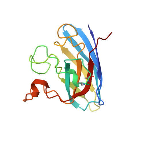
Crystal structure of reduced bovine erythrocyte superoxide dismutase at 1.9 A resolution.
Rypniewski, W.R., Mangani, S., Bruni, B., Orioli, P.L., Casati, M., Wilson, K.S.(1995) J Mol Biol 251: 282-296
- PubMed: 7643403
- DOI: https://doi.org/10.1006/jmbi.1995.0434
- Primary Citation of Related Structures:
1SXA, 1SXB, 1SXC - PubMed Abstract:
Cu,Zn superoxide dismutase was investigated crystallographically in the reduced form. Co-ordinate errors were estimated by comparing two independently refined models, based on two different data sets. This gave a detailed error estimation as opposed to the standard sigma A and Luzzati plots, which estimate only the overall error. The high quality of the final model, obtained after scaling together the two data sets, combined with the error estimates allowed a detailed analysis of the protein and solvent structures. An automatic procedure for building and refining solvent structure was tested and found to give reproducible results. Contrary to results obtained from spectroscopic studies, the co-ordination of the metal ions in the catalytic site is preserved in the crystal structure of the reduced enzyme, as compared with the crystal structure of the oxidised form. Analysis of the solvent reveals a well-defined chain of closely packed, hydrogen-bonded water molecules filling the active site groove. This structural feature could serve as a hydrogen bond relay for efficient delivery of protons to the active centre. Analysis of electron density suggests that Glu119 is covalently modified. The modification, if originated in vivo, could have a role in the catalytic mechanism and could affect the overall electrostatic field in the active site. There are significant differences between the active sites of the two crystallographically independent monomers. They are explained in terms of local differences in the crystal environment.
Organizational Affiliation:
European Molecular Biology Laboratory (EMBL), Hamburg, Germany.